Foot surgery has evolved towards minimally invasive techniques that offer great benefits over traditional procedures. At Clínica San Román, we are specialists in percutaneous foot surgery, an advanced method that allows us to treat deformities such as bunions, claw toes or hammer toes through minimal incisions and with a faster recovery.
What is foot surgery?
Foot surgery is a set of surgical techniques that correct bone and joint problems that affect mobility, comfort and general health. One of the most innovative is percutaneous surgery, which uses incisions of a few millimeters under local anesthesia and radiological control to achieve precise results with minimal aggression to the tissues.
Most common types of foot surgery
- Bunion surgery (Hallux Valgus): correction of the deviated big toe.
- Metatarsal surgery: indicated in cases of metatarsal pain or overload.
- Claw or hammer toe surgery: to improve the alignment and functionality of the fingers.
- Arthroscopic foot surgery: used to treat joints and internal tissues.
- Percutaneous surgery: the minimally invasive technique that reduces recovery time.
Advantages of minimally invasive foot surgery
- Faster recovery: allows walking soon after surgery.
- Less pain: due to small incisions and minimal tissue involvement.
- No visible scars: the incisions of only millimeters leave almost imperceptible marks.
- Fewer complications: reduces the risk of infections and bleeding.
How is the process of foot surgery
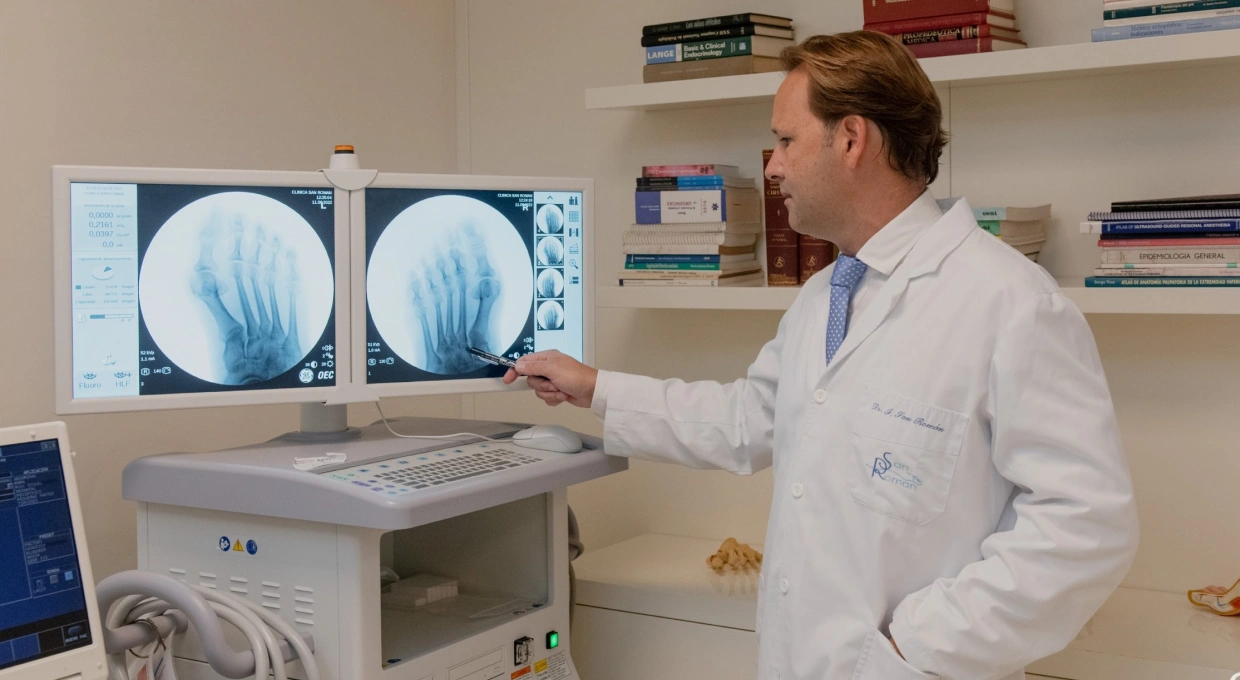
- Initial evaluation: personalized diagnosis with imaging tests.
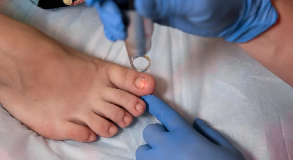
- Surgical intervention: minimal incisions guided by fluoroscopy are made to correct the deformity.
- Recovery and follow-up: the patient wears a post-surgical shoe and attends scheduled check-ups.
Recovery after foot surgery
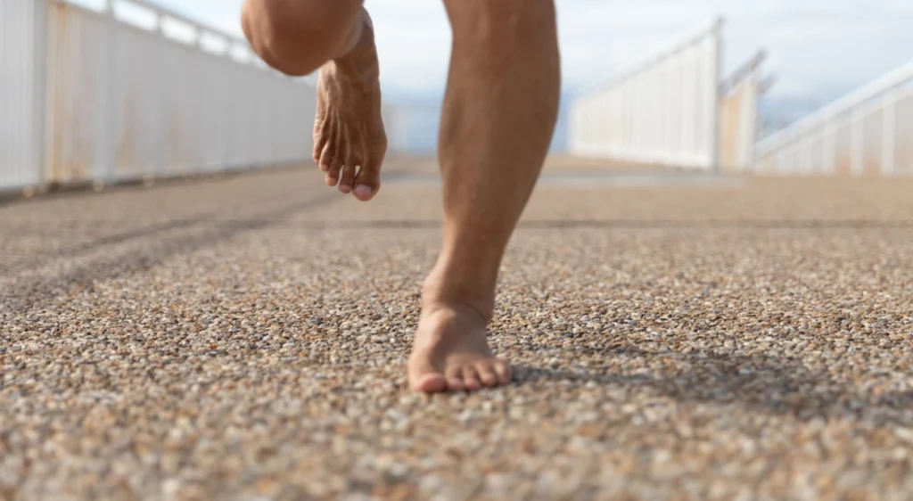
One of the main advantages of percutaneous surgery is the rapid recovery. In most cases, the patient can walk the same day of the intervention with special footwear. Swelling decreases progressively and the return to normal life is achieved in a few weeks.
- Postoperative care: keep the foot in relative rest and follow medical indications.
- Decrease inflammation: with local cold and elevation of the foot.
- Medical follow-up: periodic check-ups to ensure the correct evolution.
Advanced instrumentation and technology
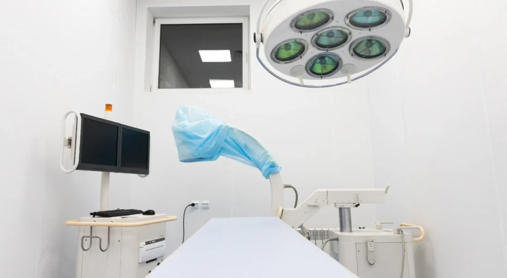
At Clínica San Román we use state-of-the-art instruments that allow us to perform foot surgeries with maximum precision and safety:
- Fluoroscope: real-time images that guide the surgeon during the operation.
- Percutaneous scalpel: designed to make minimal incisions without damaging tissues.
- Surgical drills: remodel the affected bones with high precision.
- 4D ultrasound: allows evaluation of the foot before and after bunion surgery.
Success stories at Clinica San Roman
Dozens of patients have already improved their quality of life thanks to the minimally invasive foot surgery performed in our clinic. Most of them highlight the reduction of pain, the quick recovery and the possibility of returning to their daily routine soon.
Frequently asked questions about foot surgery
- How long does the surgery last? Generally between 30 and 60 minutes.
- Does it hurt after the operation? Pain is minimal due to the percutaneous technique.
- When can I walk? Same day with post-surgical shoe.
- How long does it take for the swelling to go down? Usually between 2 and 4 weeks.
Conclusion
Minimally invasive foot surgery is a very suitable option to correct deformities effectively, with less pain and accelerated recovery. At Clínica San Román we have leading specialists in percutaneous surgery, advanced technology and the necessary experience to promote optimal and lasting results.