
MIS SURGERY – TARSAL TUNNEL SYNDROME – ALICANTE
Treatment of tarsal tunnel syndrome: we release the nerve without opening the ankle.
Percutaneous MIS decompression of the posterior tibial nerve – Local anesthesia, no plaster, same day discharge – 45 years treating foot neuropathies in Alicante.
Backed by 45 years of experience
Clinic founded in 1979
Four and a half decades treating nerve and biomechanical foot pathologies in Alicante.
Certification MIS23BE03 ABMSP
European pioneers in minimally invasive surgery of the foot accredited by the American Board of Multispecialty Surgical Podiatry.
Multilingual service
We attend you in Spanish, English, German, French and Dutch.
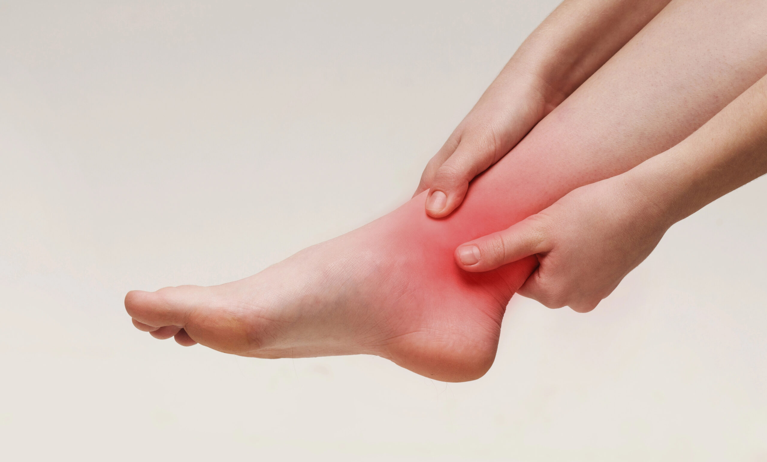
What is tarsal tunnel syndrome?
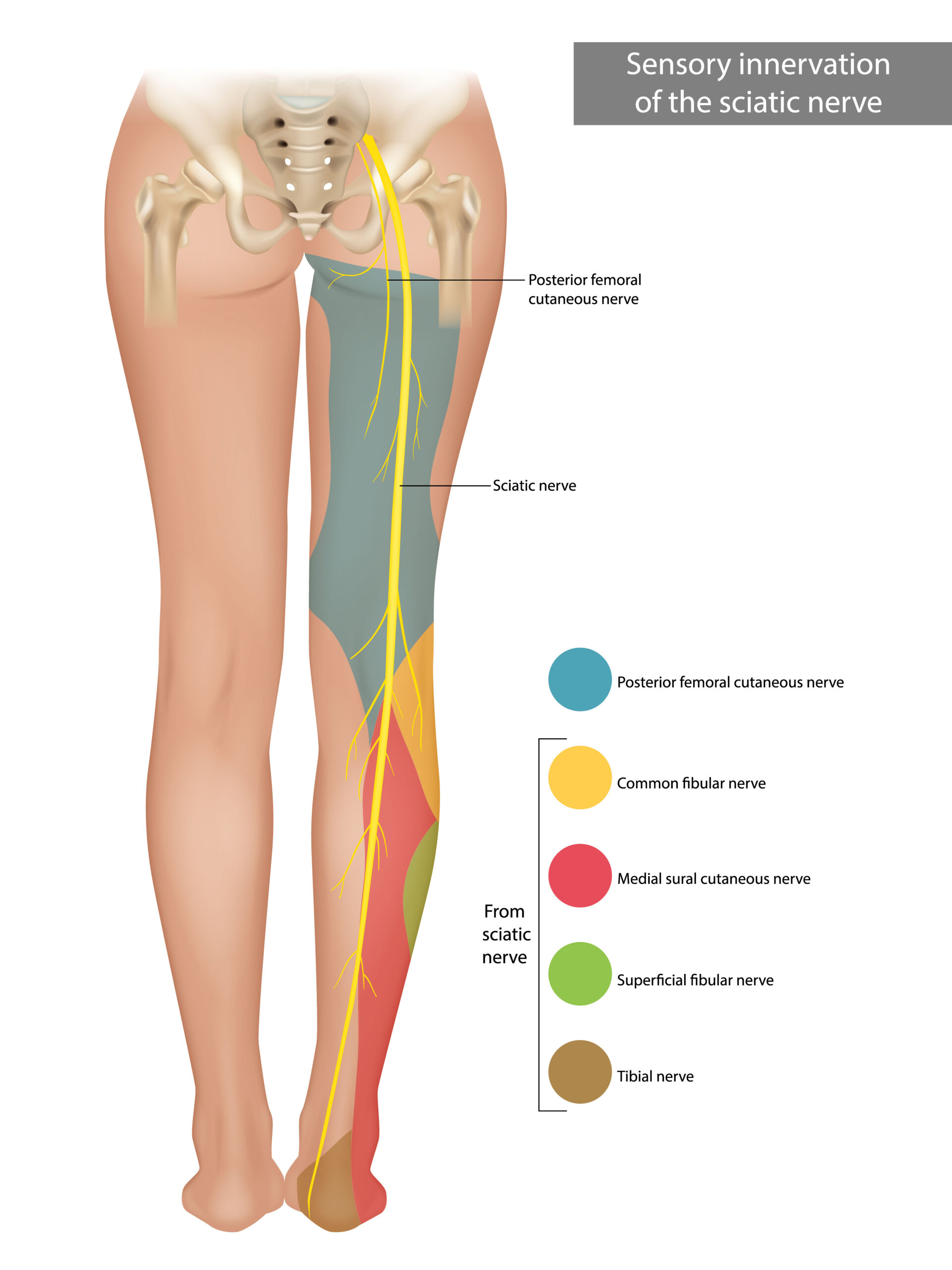
Tarsal tunnel syndrome is a compressive neuropathy: the posterior tibial nerve is trapped as it passes through an osteofibrous canal located behind and below the medial malleolus (the inner ankle bone). That channel is covered by a fibrous roof, the flexor retinaculum, which holds the flexor tendons in place. When the pressure inside the tunnel rises – due to a varicose vein, a ganglion, synovitis or sustained overpronation – the nerve becomes irritated, stops conducting the signal well and the typical symptoms of tingling, burning and loss of sensation in the sole of the foot appear.
Although often compared to carpal tunnel at the wrist, they are distinct conditions: tarsal tunnel is less commonly diagnosed and often arrives at the office later, delaying treatment. Several studies point out that the actual prevalence is underestimated because many patients are mislabeled as plantar fasciitis or posterior tibial tendinitis before the correct diagnosis is made (Lau JT, Daniels TR. Foot Ankle Int. 1999).
At Clínica San Román we approach this pathology from its root: we confirm compression with examination + dynamic ultrasound + electroneurography, we exhaust conservative options whenever indicated and, when the nerve has to be released, we do it with percutaneous MIS technique instead of traditional open surgery.
Symptoms: How to recognize a tarsal tunnel?
The classic picture is a burning or electrical sensation in the sole of the foot and toes, which many patients describe as “walking on coals”, “pins and needles” or “sleepy foot”. It is typically worse at night when getting into bed and improves when moving the foot or getting up to walk for a while. The distribution follows the territory of the posterior tibial nerve and its medial plantar, lateral plantar and medial calcaneal branches.
- Burning, tingling or cramping in the sole of the foot, toes and heel area.
- Pain that worsens at night and with prolonged standing or walking.
- Loss of sensation in the sole or sensation of “walking on cotton”.
- Positive Tinel’s sign: percussion behind the medial malleolus reproduces the electrical discharge towards the fingers.
- Weakness of the intrinsic muscles of the foot, difficulty in opening the toes.
- Pain radiating towards the calf in some cases (retrograde irradiation).
- Worsening with closed shoes, rigid heels or prolonged sports activity.
- Bilateral symptoms in a minority of patients, especially in severe pronator feet.
Why does tarsal tunnel syndrome appear?
There is no single cause: tarsal tunnel is a multifactorial condition. In about half of the cases an “occupying” cause is identified (something compressing the nerve within the tunnel) and in the rest biomechanical and systemic factors coexist. Therefore, the first step is to understand what is causing the compression in your case.
🦴 Compression due to occupying structures
Ganglions (synovial cysts protruding from the joint), varicose veins of the posterior tibial vascular bundle, synovitis of the flexor tendons, bony exostoses or sequelae of fracture of the malleolus or talus. In these cases the nerve is “cornered” by a mass that occupies part of the tunnel. Mondelli et al. (2008) describe occupying structures in about 30-40% of surgical series.
🦶 Valgus flatfoot and overpronation
When the hindfoot collapses into valgus and the internal arch drops, the tarsal tunnel is stretched and the posterior tibial nerve is tractioned against the roof of the retinaculum. This sustained traction is one of the most common biomechanical causes and the one that benefits most from conservative treatment with custom insoles and gait retraining.
🩺 Systemic and metabolic causes.
Diabetes mellitus, rheumatoid arthritis, hypothyroidism, pregnancy (due to edema and hormonal changes) and other polyneuropathies make the nerve more vulnerable to compression. In the diabetic patient the combination of background neuropathy + focal entrapment requires particularly careful management of metabolic control and surgical indication.
Diagnosis: how do we confirm tarsal tunnel syndrome?
Tarsal tunnel is diagnosed by combining a detailed clinical examination with tests that demonstrate compression and rule out other causes. In consultation we integrate the three pillars in a single visit whenever possible.
🔍 Clinical examination + Tinel
We collect the history (when it started, rate of onset, factors that worsen), assess the static and dynamic footprint and reproduce the symptoms with Tinel’s sign: by tapping the flexor retinaculum you will feel that “electric shock” to the sole of the foot. We also evaluate the sensitivity by dermatomas and the strength of the intrinsic muscles.
🔊 Dynamic ultrasound of soft tissues
It is the test that provides the highest performance in consultation. It allows to see the posterior tibial nerve in real time, to measure its cross-sectional area (a thickened nerve >0.12-0.15 cm² is very suggestive), to locate cysts, varicose veins or synovitis and to check how the nerve behaves when the foot moves. It is painless, does not radiate and is performed on the spot.
⚡ Electroneurography (EMG/ENG).
It is the reference neurophysiological test: it measures the conduction velocity of the posterior tibial nerve and its branches. It confirms entrapment and provides information on how affected the nerve is. We order it when clinical and ultrasound findings are inconclusive or when there is a need to differentiate from a baseline polyneuropathy.
Treatment of tarsal tunnel syndrome: stepwise protocol.
Our philosophy is “less is more”: we start with the least invasive and only escalate when the response is not sufficient. Surgery is an excellent tool, but we only indicate it when there is a clear structural cause or when previous treatments have failed. This is the sequence we apply.
1 – Conservative treatment
Custom insoles with hindfoot valgus correction to unload the nerve, modification of footwear (stable heel of 2-3 cm, rigid heel counter), prescribed NSAIDs, physiotherapy with neurodynamic techniques to slide and mobilize the nerve, and relative rest from the activities that trigger the symptoms. In incipient conditions or those of pure biomechanical origin, this phase resolves or significantly improves a significant proportion of patients.
2 – Echoguided infiltration with corticosteroid
When the conservative treatment is not enough and the cause is inflammatory (synovitis, perineural edema, small ganglion), we perform an ultrasound-guided infiltration with corticosteroid in the tunnel. Ultrasound allows us to deposit the drug next to the nerve without puncturing it. It works as a “pharmacological decompression” and also serves as a predictive test: if you get a lot of relief, it reinforces the diagnosis and provides guidance on the expected response to surgery if necessary.
3 – MIS Surgery: percutaneous release of the flexor retinaculum
When there is a confirmed structural cause (ganglion, varicose veins, chronic synovitis) or conservative treatment and infiltration fail to control the symptoms, MIS decompression is indicated. Under local anesthesia, a millimeter incision behind the medial malleolus and under ultrasound control, we open the fibrous roof of the tunnel and release the nerve. No screws, no plaster cast, same day discharge. Series such as Ahmad et al (2012) describe significant symptomatic improvement in most patients with identifiable structural cause.
Comparison of tarsal tunnel treatments
| Criteria | Insoles + physio | Ultrasound-guided infiltration | Percutaneous MIS surgery | Traditional open surgery |
|---|---|---|---|---|
| Indication | Mild to moderate symptoms of biomechanical origin. | Synovitis, perineural edema, initial conservative failure | Confirmed structural cause or failure of steps 1-2 | Extensive compressions, tumors, complex post-traumatic sequelae |
| Invasiveness | Null | Minimal (needle) | Minimal (millimeter incision) | High (wide incision, open dissection) |
| Anesthesia | No infiltrative | Local infiltrative | Local + optional sedation | Spinal or general |
| Sessions / time | 2-6 months of continuous use | 1-2 infiltrations every 4-6 weeks | Single outpatient intervention, ~30-45 min | Hospitalization 24-48 h, cast 2-4 weeks |
| Expected efficacy | Variable; useful in mild biomechanical forms | Relief in most in the short-medium term. | High in well-selected patients (Ahmad 2012). | High, with greater morbidity and longer sick leave |
Advantages of MIS surgery versus open surgery
👣 You walk the same day.
With post-surgical off-loading shoes you can support and return home on your own feet. No plaster cast, no mandatory crutches.
💉 Local anesthesia
We sleep only the ankle and the tunnel area. You save the risks and recovery time of general or spinal anesthesia.
✂️ Millimeter incision
The scar is practically imperceptible and minimizes the risk of nerve adhesions to the post-surgical scar tissue.
⚡ Rapid recovery
Most resume daily activities in 2-3 weeks and impact sports between the 6th and 8th week, depending on the case.
🛡️ Preserves structures
We only open the fibrous roof of the tunnel. We do not touch flexor tendons or vascular bundle beyond what is essential.
🎯 High efficiency in well selected
When there is a clear compressive cause and the nerve still conducts, release obtains consistent symptomatic improvement in the literature (Ahmad M, et al. 2012).
Prevention of tarsal tunnel
It is not always avoidable, but there are modifiable factors that reduce the risk, especially if you have valgus flatfoot or a long standing job.
👟 Suitable footwear and insoles.
Shoes with a good counter, cushioned sole and stable last. If your foot pronates, a custom insole that controls hindfoot valgus reduces the traction maintained on the posterior tibial nerve.
🤸 Mobility and proprioception.
Working on ankle mobility, strengthening the posterior tibialis and peroneals and maintaining flexibility of the Achilles tendon helps to keep the mechanics of the tunnel from being overloaded.
⚖️ Weight control and systemic pathology
Maintaining a healthy weight decreases the load on the hindfoot. If you have diabetes, rheumatoid arthritis or hypothyroidism, strict metabolic control decreases the vulnerability of the nerve.

Do you feel tingling or burning in the sole of your foot at night?
It may be your tibial nerve asking for space. We will evaluate you without obligation and explain what options you have in your case.
Request your free evaluationFrequently asked questions about tarsal tunnel syndrome
We solve the most common doubts of our patients.
🔬 About the pathology.
Is tarsal tunnel the same as carpal tunnel but in the foot?
The concept is similar – a nerve that is compressed as it passes through a canal – but neither the nerve, nor the anatomy, nor the treatment are the same. In carpal tunnel, the median nerve is trapped in the wrist; in tarsal tunnel, the posterior tibial nerve is trapped in the ankle. Tarsal tunnel is less frequently diagnosed and usually has a biomechanical component (valgus flatfoot) that is not present in the wrist. It should therefore be evaluated specifically.
Why are the symptoms worse at night?
At night, the venous return of the foot decreases and the small vessels of the tunnel ingurgitate, increasing the pressure on the nerve. In addition, by being immobile, you lose the natural “massage” that walking produces and the nerve is more exposed to the edema accumulated during the day. It is very common for patients to tell us that they get up and walk around the house to relieve themselves: this gesture momentarily reduces the intratunnel pressure.
Can it be cured without surgery?
In mild cases or those of pure biomechanical origin – pronator foot, work overload, fluctuating edema – conservative treatment with insoles, modification of footwear, physiotherapy and, if necessary, ultrasound-guided infiltration can control symptoms in a significant proportion of patients. When there is an occupying structural cause (ganglion, varicose veins, chronic synovitis) or the nerve already shows neurophysiological alteration, surgery is usually the most effective option in the medium term.
Does diabetes worsen tarsal tunnel?
Yes, diabetes makes the nerve more vulnerable to compression and, if blood glucose is poorly controlled, symptoms tend to be more intense and recovery slower. In diabetic patients, diffuse polyneuropathy and focal tunnel entrapment often coexist: this is why neurophysiological studies are so important to differentiate between the two components and to plan the treatment well, especially if surgery is considered.
🏥 Treatment
How long does the operation last?
MIS decompression of the flexor retinaculum takes 30 to 45 minutes in most cases. The previous preparation, local anesthesia and the immediate postoperative period add approximately one hour more in the clinic. It is an outpatient procedure: you walk in on your foot and walk out too, with post-surgical off-loading shoes, without the need for hospitalization or general anesthesia.
Do I walk again the same day?
Yes, the philosophy of MIS surgery is precisely that: local anesthesia, no plaster and immediate loading with post-surgical footwear. Unless medically contraindicated, you leave the clinic walking. During the first 7-10 days we recommend limiting long walks, keeping the foot elevated at rest and applying the care guidelines we give you in writing. The mobility of the ankle recovers progressively.
When do I regain full sensitivity?
Pain and burning relief is usually noticeable within the first few weeks. The recovery of fine sensation depends on how long the nerve has been compressed: if it is recent, it recovers fairly quickly (weeks to months); if you have had symptoms for years, the nerve may take 6 to 12 months to regenerate and, in some cases, a small area of altered sensation remains. We will follow up with you periodically to evaluate it.
🏃 Recovery and prevention
Can it recur after surgery?
Recurrence is possible, especially if the underlying cause has not been treated: for example, a valgus flatfoot that continues to tract the nerve or a varicose vein that grows back. Therefore, in addition to freeing the tunnel, we plan the postoperative period with corrective insoles and, if necessary, address the structural cause. Review series place recurrence in a minority of patients when the surgical indication is precise.
Can I prevent it with insoles?
If your foot pronates or you have hindfoot valgus, a custom insole that corrects the mechanics reduces sustained traction on the posterior tibial nerve. It does not guarantee that you will not develop the condition, but it clearly lowers the risk and, in patients with initial symptoms, it can slow progression. The generic insole is no good: it needs a biomechanical study and individualized design for your foot.
Scientific references
- Lau JT, Daniels TR. Tarsal tunnel syndrome: a review of the literature. Foot Ankle Int. 1999;20(3):201-9. [PMID: 10195299]
- Mondelli M, Morana P, Padua L, Reale F, Giannini F. Tarsal tunnel syndrome: clinical, electrophysiological and imaging features in 187 patients. Clin Neurophysiol. 2008;119(8):1818-1825. [PMID: 18495524]
- Ahmad M, Tsang K, Mackenney PJ, Adedapo AO. Tarsal tunnel syndrome: a literature review. Foot Ankle Surg. 2012;18(3):149-52. [PMID: 22857953]
- Kinoshita M, Okuda R, Morikawa J, Jotoku T, Abe M. The dorsiflexion-eversion test for diagnosis of tarsal tunnel syndrome. J Bone Joint Surg Am. 2001;83(12):1835-9. [PMID: 11741063]
- Antoniadis G, Scheglmann K. Posterior tarsal tunnel syndrome: diagnosis and treatment. Dtsch Arztebl Int. 2008;105(45):776-81. [PMID: 19578413]
- Fortier LM, Markel J, Thomas BG, Sherman WF, Thomas LC, Kaye AD. An update on posterior tarsal tunnel syndrome. Orthop Rev (Pavia). 2022;14(3):35444. [PMID: 35965974]
This page is for information purposes only and does not replace the individual clinical assessment. Each case requires a personalized examination.
Don’t let tarsal tunnel control your nights and your day-to-day life.
If the bottom of your foot burns, tingles or wakes you up at night, we can help. We’ll assess you, explain what’s going on and come up with a customized plan – no pressure, no empty promises.
- 📞 +34 965 921 156
- ✉️ info@clinicasanroman.com
- Av. del Dr. Ramón y Cajal 1, 03001 Alicante