Foot Surgery: Do you have pain in your feet? Minimally invasive

21 de September de 2018

Percutaneous Foot Surgery

If you have bunions, claw toes, hammer toes, or other foot deformities but are fearful of traditional surgery because of its potential risks and complex post-operative care, this information about percutaneous foot surgery at Clínica San Román may be helpful.

cirugía-percutanea-del-pie

In simple terms, percutaneous foot surgery is a technique that allows correction of severe and painful deformities such as bunions, claw toes and hammertoes through millimetre incisions without the use of pins, screws or plates.

Current trends in surgery involve performing increasingly less invasive procedures through smaller incisions because they reduce postoperative complications and shorten the recovery period. Nowadays, minimally invasive foot surgery allows good correction of foot deformities with optimal results. To successfully carry out these techniques, it is essential to have specific instruments, modern radiological equipment (fluoroscopes) and extensive surgical experience in percutaneous surgery and must always be performed by qualified surgeons.

As with other foot surgery techniques, the goal of percutaneous surgery is to reduce the deformity, eliminate pain and correct the pathological elements that caused the deformity in order to prevent recurrence while maintaining optimal function of the foot. Foot surgery is not indicated for aesthetic purposes only; each patient should be treated individually based on their circumstances and background.

For this reason, a careful clinical examination of the foot should be carried out to assess for any asymmetries, misalignment, joint alterations, foot type, etc.

At the same time, the presence of other pathologies in the patient should be assessed as these may, in turn, aggravate the presenting foot deformity. Such pathologies may include gout (high uric acid and joint pain), rheumatoid arthritis and psoriatic arthritis, among others. Moreover, the assessment should also include a study of the neurological and vascular status of the foot, especially in diabetic patients.

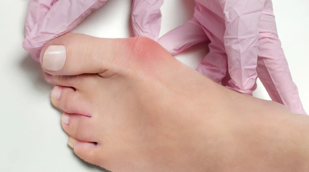
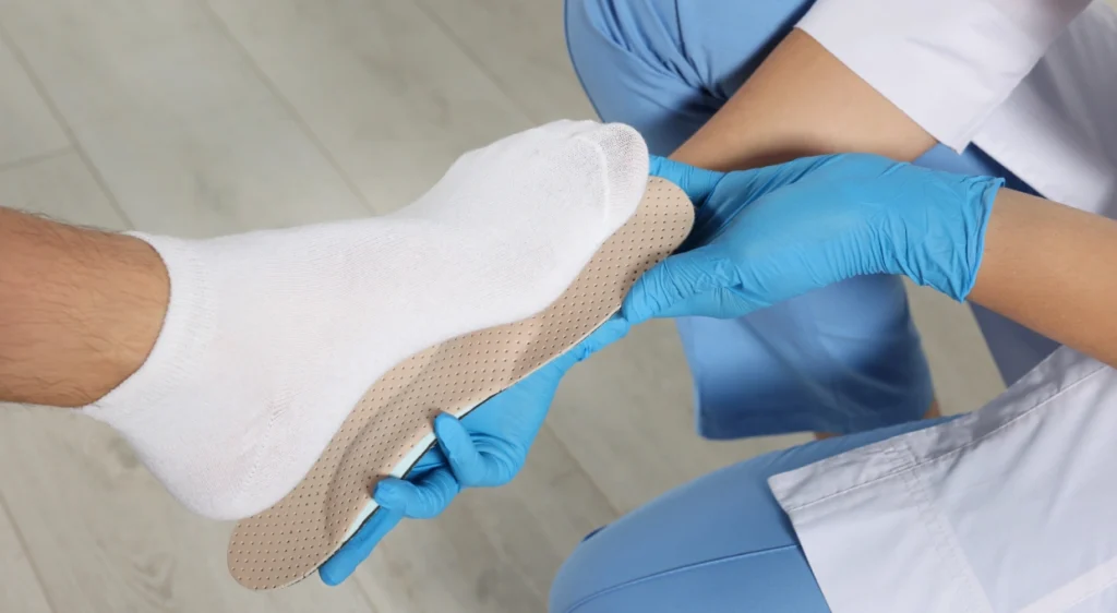
Treatment of hallux valgus may vary depending on the degree of deformity. In early deformities, conservative treatments can be performed with appropriate footwear and orthotics to slow the development of the deformity. However, the only definitive solution is surgery to remove the bony bump and correct the deviation caused by hallux valgus.

There are over 150 surgical techniques for the treatment of hallux valgus, varying from different osteotomies of the first ray, to arthrodesis with plates or screws, but there is no doubt that percutaneous surgery is the surgical technique that most respects the foot, causing minimal trauma and with fewer post-operative complications.

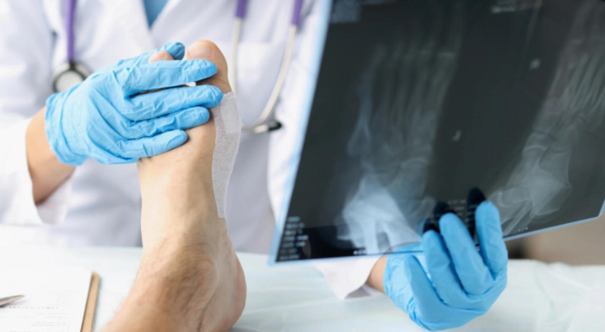
To carry out a bunion procedure using minimally invasive techniques, it is essential to use a fluoroscope that reproduce images of the foot in real time to guide the surgeon throughout the different surgical procedures.


Radiological control instruments


radiografia-del-pie


Low-intensity X-ray equipment (fluoroscope) is essential to accurately monitor and perform osteotomies (surgical bone fractures) and bone resections. When making an incision of a few millimeters, the fluoroscope is necessary because the surgeon does not have a direct view of the surgical field.

The surgery is performed on an outpatient day-case basis under local anesthesia, which reduces the possible complications and adverse effects of general anesthesia.


Bunions, claw or hammer toes

before foot surgery

Bunions, claw or hammer toes, as well as bunions, mostly affect the female sex. If you have any of these deformities, they are likely due to inheritance and/or repeated use of constrictive shoes with excessive heels.

Regardless of the cause it is important to be aware of these deformities; not only they are an aesthetic issue but also because they can have serious consequences in future deformity progression, such as dislocation of joints and increasingly large deformities that can lead to ulceration and further disability.


Minimally invasive surgery is also used for the treatment of:

  • Tailor’s bunion: this is similar to the bunion but is the bony protrusion affecting the fifth toe (fifth metatarsal head).
  • Hallux Rigidus: osteoarthritis that affects the big toe joint and produces joint stiffness and pain.
  • Calcaneal Spur: a bony prominence that occurs in the lower part of the heel that can cause pain.
  • Morton’s neuroma: A thickening of the interdigital nerve primarily in the interdigital space between the third and fourth toes.


Who is MIS surgery aimed at?

Anyone with foot deformities who has pain or cannot walk properly or wear normal shoes may benefit from these techniques.

Preparing for Percutaneous Foot Surgery:

Before the surgery is performed, the specialist must evaluate the degree of deformity to determine the best percutaneous technique to be performed.

Once the surgical technique has been decided, it is essential to perform a blood test with coagulation tests.


What does the operation consist of?

This is an outpatient day-case procedure with a local anaesthetic block that allows the area to be operated on to be anaesthetised. One or more incisions of a few millimeters are made, through which surgical procedures are performed to solve the problem and usually can be:


Complete resection of the bony bump.

Depending on the degree of deformity, an osteotomy, i.e. a cut or partial section of the bone, is performed to recover the original position.

Release of the joint capsule or tendon, i.e. the abductor tendon of the first toe.

Correction of associated deformities, such as claw or hammer toes through 2 mm incisions.


These surgical procedures are performed under fluoroscopy guidance with real-time images of the inside of the foot.


There are more than 150 surgical techniques for the correction of these deformities, but without a doubt the interventions with percutaneous foot surgery offer the following advantages:

The incisions are only of a few millimeters, with less trauma to the foot and therefore better and faster postoperative recovery.

No surgical screws or pins are used, thus reducing possible infection problems.

The anesthesia is local, thus reducing the risks involved in general or general anesthesia.

After surgery, you walk on your own foot with a post-surgical shoe without the need of complete rest that is usually necessary with other traditional surgical procedures.

In general, percutaneous surgery allows for a very quick recovery with fewer complications compared to other more aggressive techniques.

Of course, there may always be possible complications with any foot surgery or in the postoperative period, so it is very important that this technique is performed by very experienced professionals in optimal conditions.


Procedure aftercare:

 foot surgery bunion

After the operation, the patient will have to wear the post-surgical shoe for one to five weeks to be able to walk safely.


The first check-up is carried out after 7 days to remove the stitches. Afterwards, the surgical dressing will need to be changed approximately every 14 days until surgical discharge. Finally, in order to carry out the daily cleaning of the foot, you will be provided with specially designed foot covers.

San Román Podiatric Clinic

If you are interested in this article, please contact the specialists in percutaneous or minimal incision surgery at Clínica San Román, both doctors and podiatrists who will evaluate your problem with the utmost professionalism and provide you with the best solution.

Remember that the feet are the fundamental pillar that must support your body throughout your life.

Encourage yourself to end the pain and improve your quality of life!


sello-informacion-medica-de-confianza-clinica-san-romanArticle prepared by Clínica San Román

Date of Publication: 21-09-2018


Share

Related posts