ADVANCED PODIATRY · FREIBERG’S DISEASE · ALICANTE
Freiberg’s Disease: Diagnosis and Specialized Treatment
Avascular necrosis of the metatarsal. Early diagnosis. Conservative and surgical treatment adapted to each stage.
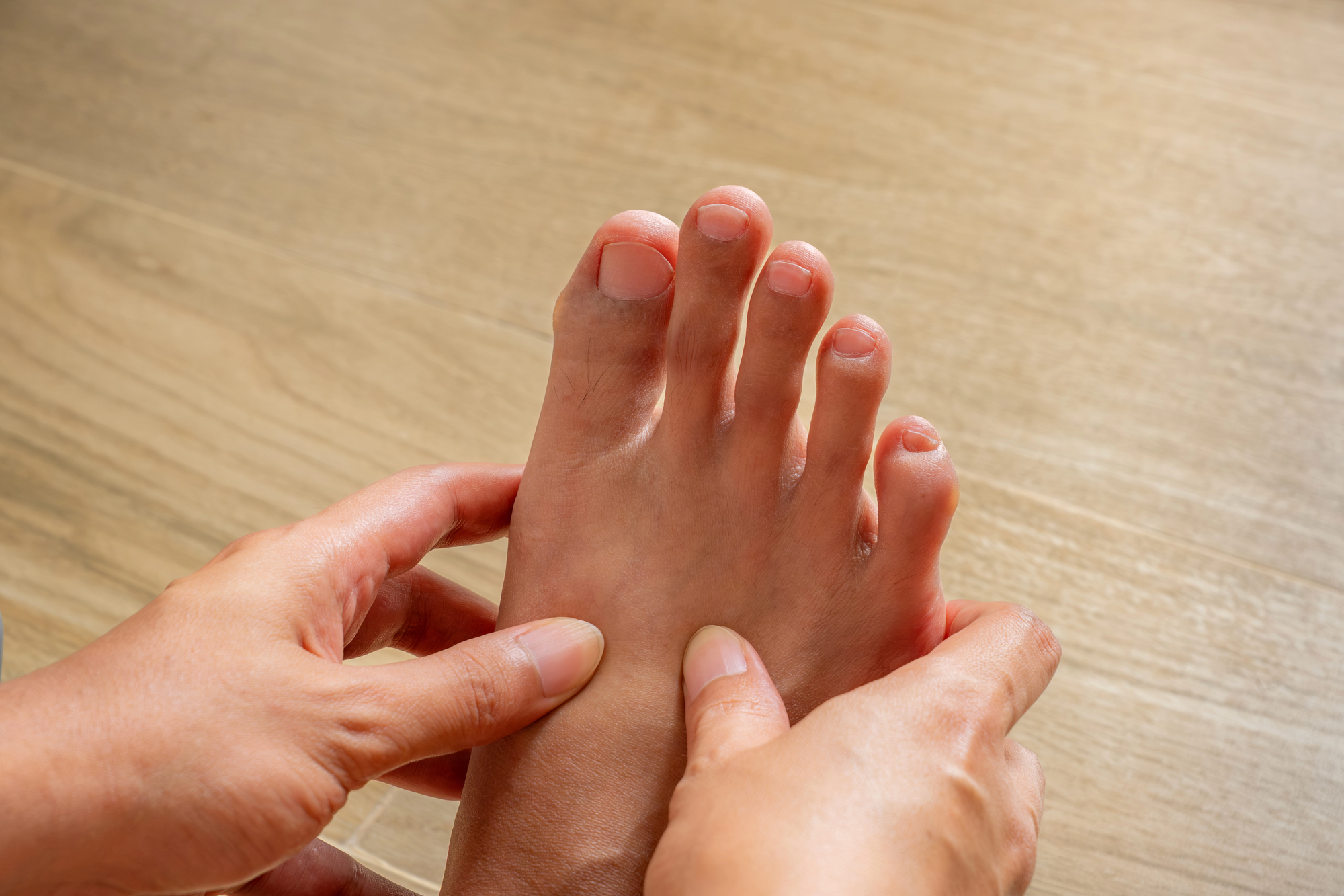
If you’ve been experiencing pain under your second toe when walking for months, or if you notice that your foot is swollen and the joint has become stiff—you may be suffering from Freiberg’s disease. It is a little-known condition, but one with serious consequences if not treated promptly. At Clínica San Román, we have over 45 years of experience in foot conditions and offer a personalized approach based on the stage of the disease: from orthotics and weight-bearing relief to percutaneous surgery under local anesthesia.
⭐ 4.8/5 (190 reviews) · 🏅 Cert. MIS23BE03 · 🇪🇺 5 languages
Backed by 45 years of experience
45+ years old
Family clinic founded in 1979
4.8 ★
190 reviews on Google
MIS 🏅
First in Europe to offer an MIS subspecialty (Cert. MIS23BE03)
🇪🇺 5
Languages: ES · EN · DE · FR · NL
What is Freiberg’s disease?
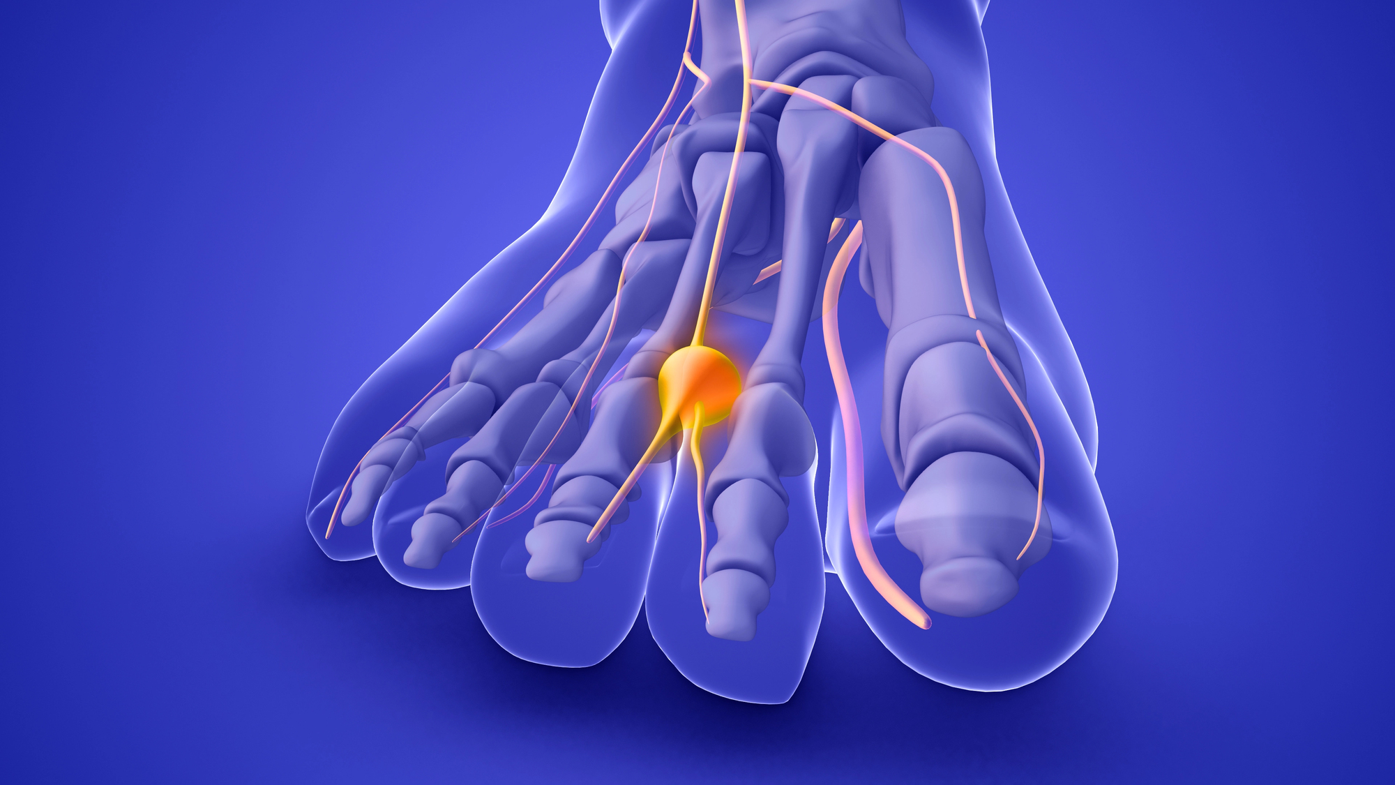
Freiberg’s disease (also known as Freiberg’s syndrome or osteochondritis dissecans of the metatarsal) is avascular necrosis of the head of the second metatarsal bone in the foot. Simply put: the blood supply to the bone in that area is cut off, and part of the bone “dies” and progressively deforms.
It was first described by Dr. Albert Freiberg in 1914. It primarily affects the second metatarsal (in 68% of cases), although it can also occur in the third (27%) and even the fourth. It is bilateral (affecting both feet) in approximately 6–10% of patients.
Although it can occur at any age, it is particularly common in adolescent and young women (ages 13–18), with a ratio of 5 women to every man. However, many cases are not diagnosed until adulthood, when the joint deformity is already advanced and chronic pain sets in. Early diagnosis is key to preventing progression to metatarsophalangeal osteoarthritis.
Symptoms and classification: How does the disease progress?
The main symptom is pain located beneath the head of the second metatarsal bone (the ball of the foot) that worsens when walking, running, or wearing high heels. Other common signs include:
- Swelling and stiffness in the second metatarsophalangeal joint — the toe moves less than usual
- A palpable joint swelling felt upon examination of the top of the foot
- Progressive limp — the patient unconsciously changes their gait to avoid pain
- Pain when bending the finger upward (dorsiflexion) or when squeezing it from the side
The disease is classified into five stages according to Smillie, which determine its severity and the most appropriate treatment:
- Stage I — Stress fracture: fissure in the ischemic epiphysis. X-ray showing increased bone density. No visible deformity.
- Stage II — Central resorption: resorption of the trabecular bone. The dorsal articular surface begins to sink slightly.
- Stage III — Collapse with bony spurs: the joint surface collapses further. Lateral bony spurs appear, visible on X-rays.
- Stage IV — Loose bodies: osteochondral fragments break off and migrate toward the joint. Significant deformity of the metatarsal head.
- Stage V — Osteoarthritis: complete deformity with flattening of the metatarsal head, osteophytes, and advanced joint degeneration.
💡 Did you know that…? Many patients with stage I–II Freiberg’s disease experience little to no discomfort, and the condition is often discovered incidentally on an X-ray. However, the earlier it is detected, the better the treatment outcomes. If you have persistent pain in the ball of your foot, consult a specialist.
What causes Freiberg’s disease?
The exact cause is not fully understood, but current scientific evidence points to a combination of factors that disrupt blood flow to the head of the metatarsal bone:
🦔 Anatomical factors
- Long second metatarsal — a metatarsal that is longer than the first (Greek metatarsal formula) bears a greater load and is more susceptible to ischemia
- Insufficient first ray — if the first metatarsal is short or hypermobile, the load is transferred to the second
- Vascular characteristics — the head of the second metatarsal bone has a single terminal blood supply (a single dominant artery), which makes it particularly vulnerable
⚡ Mechanical factors
- Repetitive microtrauma — repeated impacts with the ground while running, jumping, or dancing (very common among ballet dancers)
- High-heeled shoes — shift body weight to the forefoot, increasing pressure on the metatarsal heads
- Sports-related overuse — high-impact sports (running, tennis, jumping) without proper footwear can trigger the condition
Essentially, Freiberg’s disease occurs when an anatomically vulnerable bone is subjected to excessive mechanical stress that exceeds its capacity for vascular repair. The result is a subclinical stress fracture followed by necrosis (death) of the bone tissue and subsequent collapse of the articular surface.
Diagnosis: How is Freiberg’s disease diagnosed?
Early diagnosis is essential to prevent the disease from progressing to stages involving irreversible joint damage. At Clínica San Román, we conduct a comprehensive evaluation that includes:
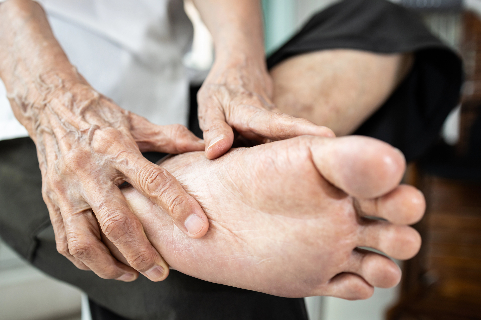
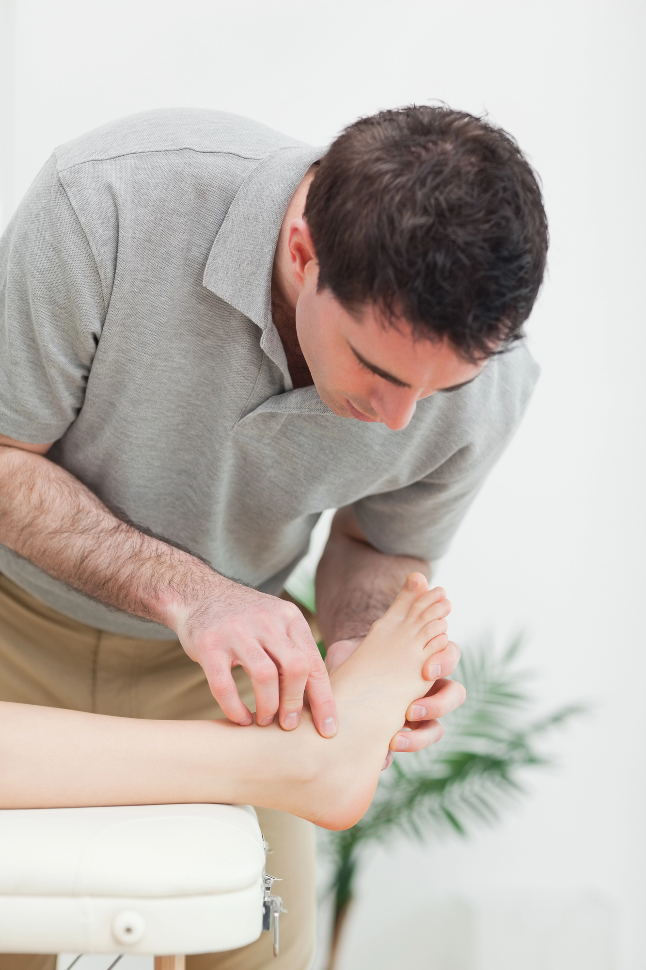
🔍 Clinical examination
Palpation of the metatarsophalangeal joint, assessment of range of motion (especially dorsiflexion), lateral compression test, and gait analysis. Joint swelling and pain on forced dorsiflexion are highly suggestive signs.
📷 Weight-bearing X-ray
Anteroposterior and lateral weight-bearing X-rays of the foot are the tests of choice. They allow for the identification of metatarsal head flattening, sclerosis, and fragmentation, and enable the classification of the disease according to Smillie’s 5-stage system.
🧲 Magnetic resonance imaging (MRI)
In the early stages, an X-ray may appear normal. An MRI detects changes in the bone marrow (edema) before deformity develops, allowing for early diagnosis and more effective conservative treatment.
Treatment of Freiberg’s disease at Clínica San Román
Our approach follows the evidence-based algorithm published in the journal Cartilage (Yoshimura et al., 2024), tailoring treatment to each patient’s stage:
Stages I–II: conservative treatment
- Custom insoles with metatarsal relief (retrocapital bar) to redistribute pressure and protect the head of the affected metatarsal bone
- Footwear modification — a rigid rocker-style sole that limits dorsiflexion of the forefoot
- Reduction of high-impact activities for 4–8 weeks, combined with gentle mobilization physical therapy
- Nonsteroidal anti-inflammatory drugs (NSAIDs) for the management of acute pain (always under a doctor’s supervision)
Stages III–V: Surgical Treatment
When conservative treatment fails to relieve symptoms (after 3–6 months) or joint deformity is significant, surgery is the most effective option. The main techniques are:
- Dorsiflexion wedge osteotomy (preferred technique) — a controlled cut is made in the head of the metatarsal bone to reposition the healthy cartilage in the weight-bearing area. Excellent results in 80% of cases with mean AOFAS scores of 96.8/100
- Joint debridement + decompressive drilling — for stage III cases with focal lesions, this procedure helps stimulate cartilage repair
- Modified Weil osteotomy (metatarsal shortening) — relieves pressure on the joint by elevating and shortening the metatarsal bone in advanced stages
- Autologous osteochondral transplantation (OAT) — in selected stage V cases, a cylinder of healthy cartilage is transferred from the knee to the head of the metatarsal bone
Treatment Options: A Comparison
Each technique has specific indications depending on the stage of the disease and the patient’s characteristics.
| Feature | Conservative (templates + download) | Dorsiflexion osteotomy | Modified Weil | OAT Transplant |
|---|---|---|---|---|
| Main indication | Stages I–II | Stages III–IV | Stages IV–V | Stage 5 selected |
| Anesthesia | Not applicable | Local | Local / regional | Regional |
| Recovery of ambulation | Immediate | Immediate with postqx footwear | 2–3 weeks | 4-6 weeks |
| Return to sports | 4–8 weeks | 2–3 months | 3-4 months | 4-6 months |
| Protects the joint | Yes | Yes | Partially | Yes (reconstructs) |
| Results (AOFAS) | Variable | 96,8/100 | 89-93/100 | 85-92/100 |
| Scar | None | Millimeter (percutaneous) | 2–3 cm | 2 cm + donor |
* Results based on published evidence (Yoshimura et al. 2024, Çakmak et al. 2016). Individual results may vary.
Advantages of the minimally invasive approach
Percutaneous surgery for Freiberg’s disease offers significant advantages over traditional open techniques:
🚶
Immediate ambulation
The patient walks in postoperative shoes starting on the day of the procedure
💈�/p>
Local anesthesia
No risks associated with general anesthesia. Ideal for young patients and athletes
🔬
Millimeter incision
Minimal tissue trauma, lower risk of infection, and faster healing
🏃
Rapid recovery
Return to sports in 2–3 months. No screws or osteosynthesis hardware
📊
Excellent results
80% excellent results. Average AOFAS score of 96.8/100 in the published evidence
🛡️
Protects the joint
Osteotomy repositions healthy cartilage, preserving long-term joint function
Do you have pain in the forefoot? See a specialist
Freiberg’s disease is a rare condition, but one that can have serious consequences if diagnosed late. Many patients live with pain and limping for years, thinking it is “normal” or simply metatarsalgia.
At Clínica San Román, Dr. Israel San Román and Dr. José Manuel San Román have some of the most extensive experience in Spain in the field of forefoot disorders. We offer a free evaluation, including an X-ray diagnosis, to determine the exact stage of your condition and recommend the most appropriate treatment.
We provide service in Spanish, English, German, French, and Dutch. Our patients travel from all over Europe to receive specialized treatment.
📍 Downtown Alicante · 📞 +34 965 921 156 · 📧 info@clinicasanroman.com
Frequently Asked Questions About Freiberg’s Disease
We solve the most common doubts of our patients. If your question isn’t listed here, please contact us and we’ll get back to you with no obligation.
📚 Scientific references (PubMed)
- Yoshimura I, Takao M, Wagner E, et al. Evidence-Based Treatment Algorithm for Freiberg Disease. Cartilage. 2024;15(1):53-63. doi:10.1177/19476035231205676. PubMed 37815268
- Çakmak MF, Bora A, Sipahioğlu S. Results of two different surgical techniques in the treatment of advanced-stage Freiberg’s disease. Indian Journal of Orthopaedics. 2016;50(2):172-177. doi:10.4103/0019-5413.173514. PubMed 27065081
- Gauthier G, Elbaz R. Freiberg’s fracture: a subchondral bone fatigue fracture. Clin Orthop Relat Res. 1979;(142):93-95. PubMed 498653
- Katcherian DA. Treatment of Freiberg’s disease. Orthop Clin North Am. 1994;25(1):69-81. PubMed 8290235
- Lee HJ, Kim JW, Min WK. Surgical treatment of Freiberg disease using extra-articular dorsal closing-wedge osteotomy: technical tips and clinical outcomes in 13 patients. Foot Ankle Int. 2013;34(1):111-116. doi:10.1177/1071100712460225. PubMed 23386770
- Al-Ashhab ME, Kandel WA, Rizk AS. A simple surgical technique for the treatment of Freiberg’s disease. The Foot. 2013;23(4):155-158. doi:10.1016/j.foot.2013.09.003. PubMed 24183700
- Carmont MR, Rees RJ, Blundell CM. Review of current concepts: Freiberg’s disease. Foot & Ankle International 2009;30(2):167-176. doi:10.3113/FAI.2009.0167. PubMed 19254514
- Smillie IS. Treatment of Freiberg’s fracture. Proc R Soc Med. 1967;60(1):29-31. PubMed 6018592
Don’t let forefoot pain limit your life
If you’ve been experiencing pain under your second toe, joint swelling, or limping for some time, don’t wait for the deformity to progress. An early diagnosis can make the difference between simple conservative treatment and more complex surgery.
San Román Clinic: Over 45 years of caring for your feet. Free consultation.