
PODIATRY – CHRONIC TALALGIA – ALICANTE
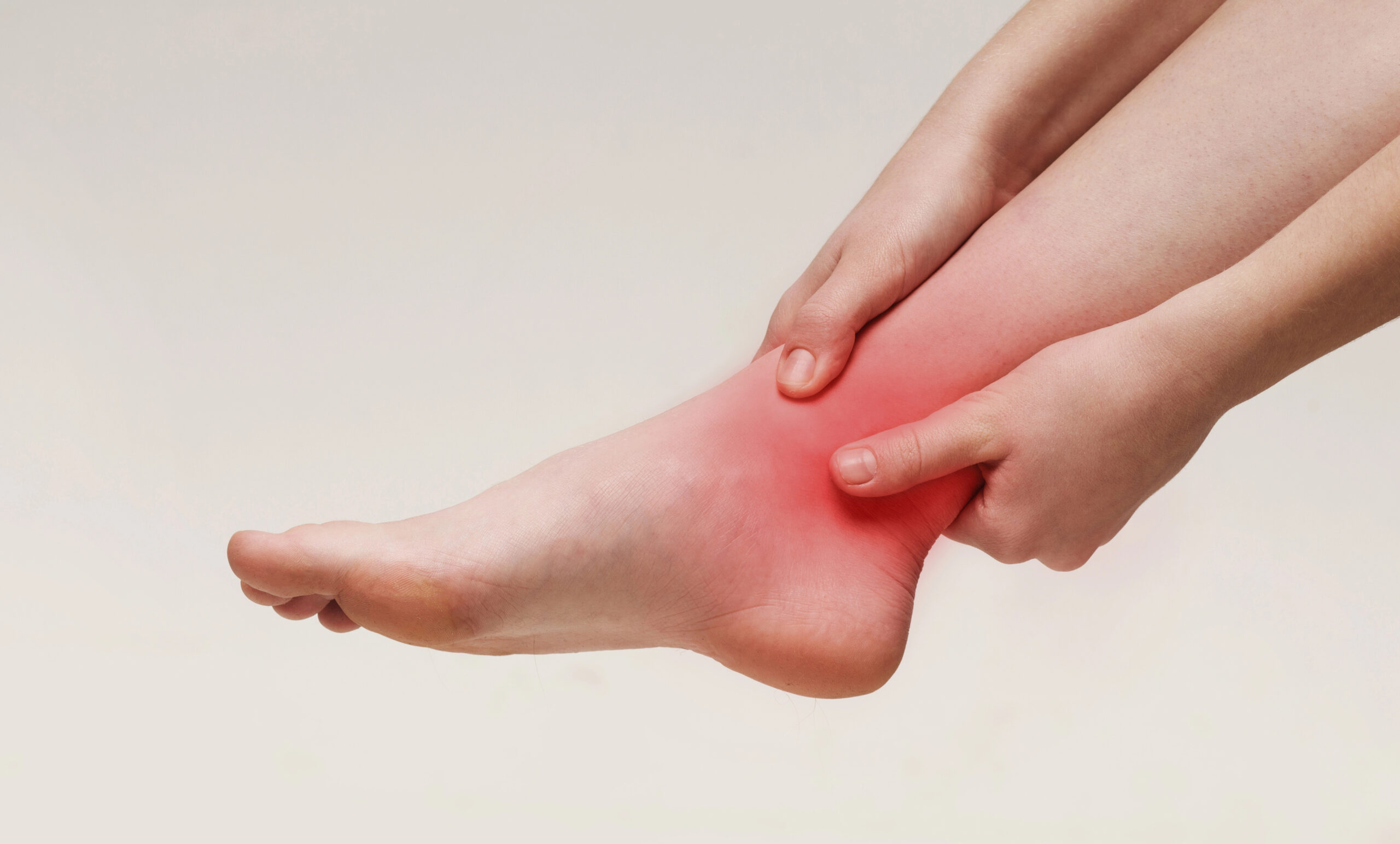
Calcaneal congestion syndrome: heel pain that X-ray does not always explain
Stepwise protocol with unloading, shock waves and ultrasound-guided infiltration. Accurate differential diagnosis of chronic heel pain. 45 years specializing in foot pathology.
45 years treating heel pain in Alicante
Clinic founded in 1979
Three generations specialized in talalgia, fasciitis and hindfoot pathology.
Certification MIS23BE03
Accredited by the American Board of Multispecialty Podiatry for minimally invasive surgery of the foot.
Multilingual service
We attend you in Spanish, English, German, French and Dutch.
What is calcaneal congestion syndrome?
Calcaneal congestion syndrome – also described in the literature as Lelièvre’s talalgia, heel pad syndrome or, in its form with established bone edema, calcaneal medullary edema syndrome –encompasses a group of conditions that share the same symptom: deep, dull and persistent pain in the sole of the heel, without major structural lesion on plain radiography.
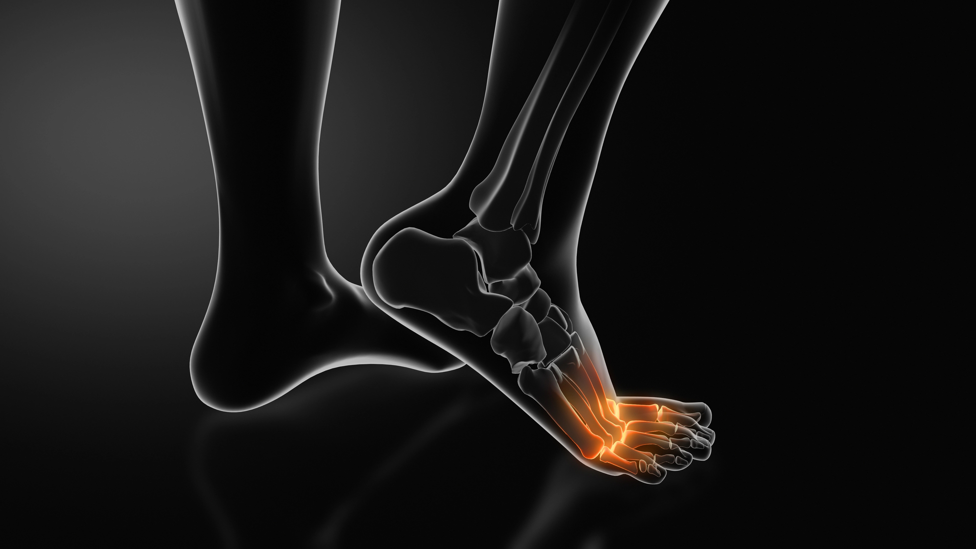
The calcaneus is the most vertically loaded bone in the human body. At each step it receives between 110% and 200% of the body weight, cushioned by a sophisticated plantar adipose bearing composed of compartmentalized fatty tissue cells that act as a biological suspension. When this structure is overloaded, atrophies or undergoes an increase in intraosseous pressure (the “congestion” described by Lelièvre in 1953), chronic pain appears which is difficult to control with simple analgesia and which radiography usually does not explain.
At Clínica San Román we approach it as a treatable mechanical-vascular condition, not as an inevitable fate: a three-level protocol – personalized unloading, shock waves and ultrasound-guided infiltration – and, only in cases with associated structural pathology (Haglund’s deformity, symptomatic spur, stress fracture), coordinated referral to trauma surgery.
Symptoms: how to recognize calcaneal congestion?
The pain of calcaneal congestion has a characteristic clinical pattern that allows it to be differentiated from plantar fasciitis and symptomatic spur. It usually develops progressively, without a single blow, and worsens with prolonged use of the foot. Most patients consult after weeks or months of evolution, when the pain begins to limit daily activity.
- Deep, dull, dull pain centered on the sole of the heel, not on the inner edge of the heel (unlike plantar fasciitis).
- Sensation of “stepping on a stone” or on a ball at the first support.
- Worsening with prolonged walking, especially on hard surfaces (asphalt, tile).
- Pain on digital pressure of the center of the heel (sensitive bearing sign).
- Partial improvement when using shoes with good cushioning or viscoelastic heel cushion.
- Burning sensation or congestion at the end of the day in prolonged episodes.
- Discrete swelling and tenderness on palpation of the heel, without marked redness.
- Absence, in most cases, of acute morning pain characteristic of plantar fasciitis.
Why does calcaneal congestion appear?
Calcaneal congestion is almost always the result of an imbalance between the load applied to the heel and the ability of the fat pad and bone to cushion it. Identifying the driving force of the condition is what differentiates a treatment that resolves from a treatment that only temporarily relieves it.
⚖️ Mechanical overload
The most frequent factor: overweight, prolonged walking on hard surfaces, static standing professions (healthcare, hospitality, retail) and sudden increases in sporting activity. The calcaneus receives repetitive loads for which its bearing was not prepared and inflammation appears. The great majority of chronic talalgias that we see in consultation have this substrate.
👟 Unsuitable footwear
Soles that are too flat and hard (flip-flops, poorly adapted minimalist sports shoes, safety shoes), excessive heel support or running shoes with depleted cushioning multiply the load on the heel. This is the reason why the picture reappears after a change of footwear or when using rigid safety shoes for the first time at work.
🦶 Predisposing biomechanics.
The cavus foot supports more vertical load on the heel. Supination gait, limb dysmetry, triceps suralis shortening and first radius stiffness selectively overload the calcaneus. Fat pad atrophy due to aging or secondary to repeated corticosteroid infiltration aggravates any overload.
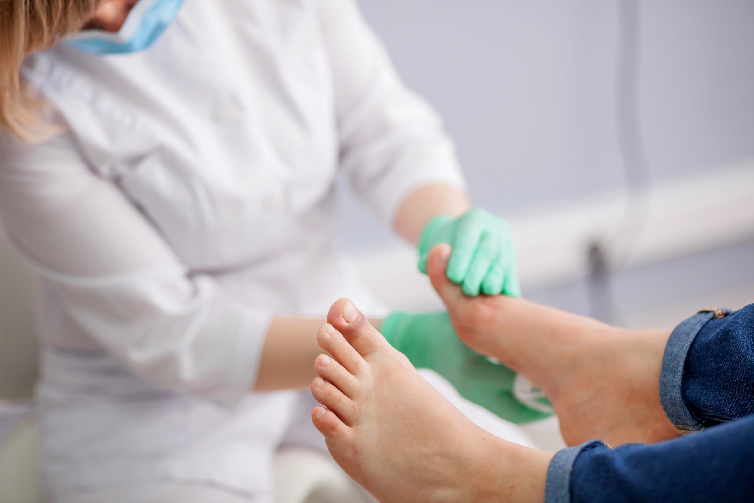
Diagnosis: how do we confirm calcaneal congestion?
A good diagnosis avoids months of failed treatments. At Clínica San Román we structure the assessment of heel pain in three complementary levels to differentiate calcaneal congestion from plantar fasciitis, symptomatic heel spur and less frequent osseous or neurological causes.
🔍 Structured clinical examination
We palpate the heel at four points: insertion of the fascia (suspected fasciitis), center of the plantar cushion (suggestive of calcaneal congestion), deep medial aspect (Baxter’s) and posterior aspect (Haglund’s, Achilles tendinopathy). We assess static and dynamic gait, ankle flexibility and possible dysmetria. A well done examination guides the diagnosis in most patients.
📷 Bilateral loading radiography
We request lateral radiography of the rearfoot in load and axial projection of the calcaneus. It allows us to assess the presence and size of the spur, Böhler’s angle, signs of stress fracture (Looser’s lines), posterosuperior Haglund’s deformity and Achilles calcifications. It is the first-line imaging test and is sufficient to guide most cases.
🔊 Ultrasound and magnetic resonance imaging
Ultrasound provides information on the fat pad (thickness measurement, cell integrity), subcalcaneal bursitis and fascia status. Magnetic resonance imaging is the gold standard when there is suspicion of calcaneal bone edema, stress fracture, fat pad injury or the need to rule out benign tumor lesions. It is reserved for cases refractory to 6-8 weeks of well-established treatment.
Treatment of calcaneal congestion: stepwise protocol
In clinical practice, calcaneal congestion syndrome is almost always resolvable without surgery. The key is not in a miraculous technique, but in applying well the combination of unloading, adjuvant therapy and infiltration when appropriate, and in correcting the biomechanical substrate that triggered it.
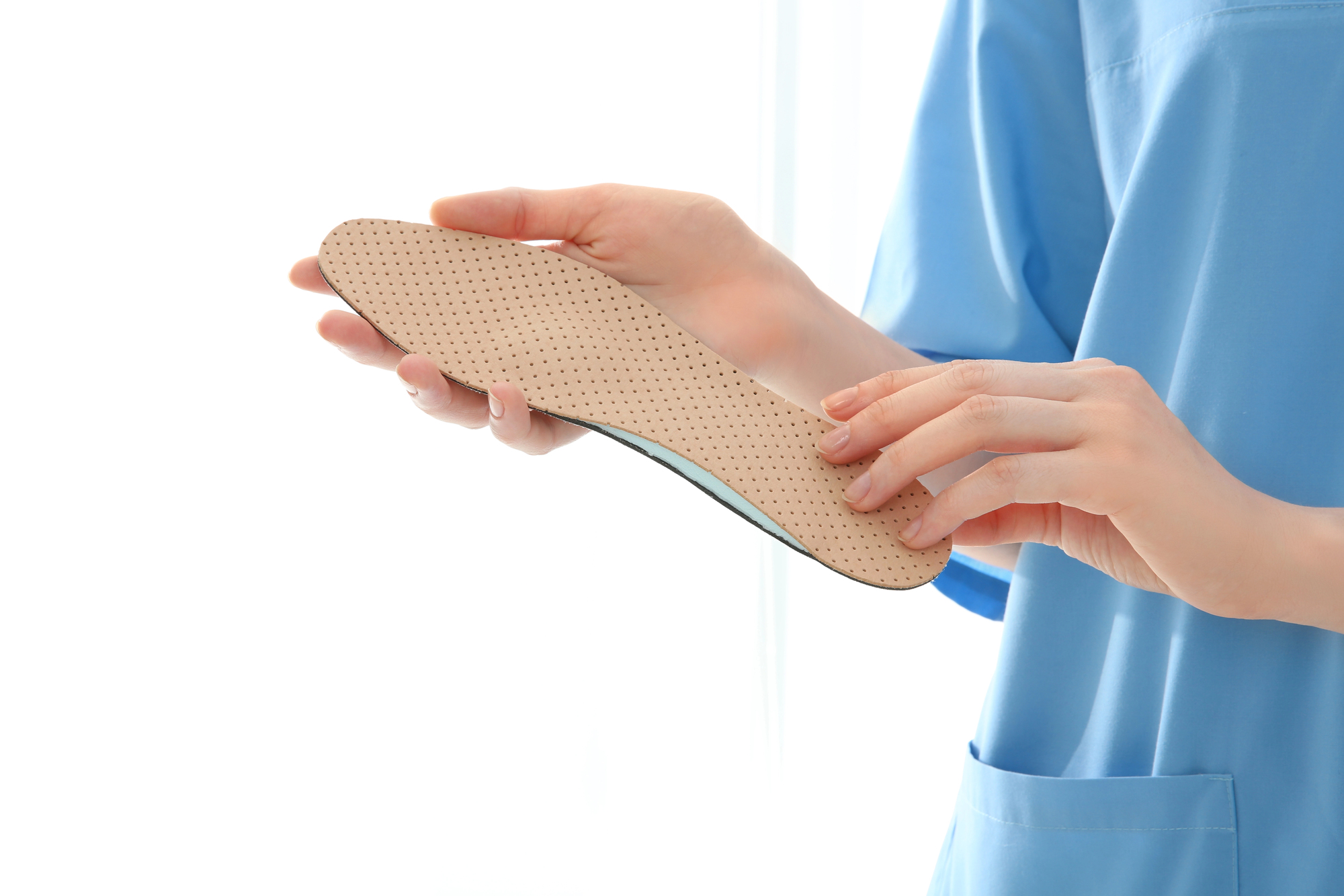
1 Conservative treatment: unloading and biomechanics
It is the basis of the protocol. It combines a quality viscoelastic heel cushion and, above all, a customized insole with heel relief designed based on gait analysis and structured exploration. We add footwear modification (sole with good rear cushioning, elimination of hard flat shoes or excessive heels), short course of oral NSAIDs (7-10 days), physiotherapy with stretching of the triceps suralis and plantar fascia, local cryotherapy and, fundamentally, adjustment of the weekly activity load. A considerable majority of patients improve in 4-6 weeks with this first line well applied.
2 Extracorporeal shock waves (ESWT)
When pain persists after 6-8 weeks of correctly performed conservative treatment, we escalate to extracorporeal shock wave therapy. It is an outpatient technique, without low labor, which uses focused acoustic waves to stimulate tissue regeneration, modulate chronic inflammation and improve local microcirculation. We recommend 3-5 sessions spaced one week apart, well tolerated. The available scientific evidence places the significant clinical response around a notable percentage of patients at 12 weeks, especially in conditions of more than 3 months of evolution (Sun et al., Foot Ankle Int 2023).
3 Selective ultrasound-guided infiltration
Indicated in cases refractory to the first two lines, especially when there is subcalcaneal bursitis or a frank inflammatory component. We perform it under ultrasound guidance, infiltrating extended release corticosteroid or platelet-rich plasma (PRP) in the peritalar space, never inside the adipose pad. We limit the number (rarely more than 2 per episode) so as not to atrophy the bearing and we always combine it with a customized insole and biomechanical adjustment. Isolated infiltration, without addressing the cause, is a patch that recurs.
4 Surgery (exceptional, in associated pathology)
Calcaneal congestion syndrome, understood as heel pain without surgical injury, is practically not operated on. Surgery is reserved for cases in which there is a structural pathology that requires surgical treatment: Haglund’s deformity with recurrent posterosuperior bursitis, symptomatic heel spur after more than 12 months of failed conservative treatment, calcaneal stress fracture that does not heal, or refractory Baxter’s nerve entrapment. In these cases we coordinate the indication with the referral orthopedic surgery and traumatology service. Percutaneous MIS surgery is an option for some of these specific indications.
Comparison of treatments for chronic heel pain
| Criteria | Template + shock | Shock waves | Ultrasound-guided infiltration | Surgery (exceptional) |
|---|---|---|---|---|
| Indication | First line, all patients | Persistent pain >6-8 weeks despite conservative treatment | Subcalcaneal bursitis / refractory after ESWT | Associated structural pathology (Haglund’s, spur, fracture) |
| Invasiveness | Null | None (without anesthesia) | Minimal (ultrasound-guided puncture) | Variable (MIS or open depending on pathology) |
| Sessions / duration | 4-6 weeks initial improvement | 3-5 weekly sessions | 1-2 sessions spaced 6 weeks apart | Multidisciplinary consultation |
| Work leave | No | No | No | Yes, according to technique |
| Evidence | Historical pillar, high adherence | High (Sun 2023; Yi 2011) | Moderate as adjuvant | Selective indication, high efficacy in the right case |
Source: Sun K et al, Foot Ankle Int 2023; Yi T-I et al, Ann Rehabil Med 2011; Mills KA et al, JOSPT 2022. Individual results may vary.
Why treat calcaneal congestion at Clínica San Román?
Accurate differential diagnosis
We differentiate congestion, fasciitis, spur, Baxter and Haglund with structured examination and imaging. Without correct diagnosis there is no effective treatment.
Advanced discharge technology
Custom insoles with viscoelastic core and selective unloading of the calcaneus from gait analysis in presoscanner.
Shock waves with evidence
ESWT equipment with validated protocol in chronic thalalalgias. Ambulatory, without sick leave, well tolerated and with documented efficacy.
Echoguided infiltration
When necessary, we always use ultrasound: millimetric precision and minimum risk of bearing atrophy.
45 years of experience
Three generations specialized in foot pathology. Surgical and conservative volume that allows fine-tuning each indication.
Multidisciplinary care
Coordination with traumatology, rheumatology and rehabilitation when required. One team, one strategy.
Prevention and maintenance
Calcaneal congestion often recurs if the measures that resolved it are not maintained. These three levers summarize the maintenance plan that we recommend at clinical discharge:
👟 Footwear and insole
Everyday shoes with good rear cushioning and removable insole (to use the customized one). In impact sports, shoe replacement every 600-800 km. Limit long days with hard flat shoes or excessive high heels. The maintenance insole is checked every 12-18 months.
🦵 Stretching and strength
Daily work of the triceps suralis, stretching of the plantar fascia, strengthening of the tibialis posterior and the intrinsic musculature of the foot. Ankle stiffness in dorsiflexion is one of the main drivers of heel overload. Five minutes a day avoids months of treatment.
⚖️ Load and weight
Control of body weight in overweight patients (every kilo counts on the heel), sporting progression without sudden jumps (10% weekly rule) and real rest between demanding days of standing. In standing professions, alternation of surfaces and unloading breaks during the day.
Have you been experiencing heel pain on heel strike for weeks?
Calcaneal congestion is effectively treated and rarely requires surgery. We study your footprint, differentiate it from other causes of heel pain and design a plan that restores the heel’s cushioning capacity.
Request your free evaluationFrequently asked questions about calcaneal congestion
We solve the most common doubts that we are asked in consultation.
🔬 About the pathology.
What is the difference between calcaneal congestion, plantar fasciitis and spur?
The pain of plantar fasciitis is localized at the inner insertion of the fascia, just at the anteromedial border of the heel, and worsens with the first steps in the morning. The heel spur is a calcification visible on X-ray that in many cases does not generate pain by itself. Calcaneal congestion produces a more diffuse, deeper pain centered on the plantar aspect of the heel, associated with overload, fat pad atrophy or increased intraosseous pressure. The three entities can coexist and share risk factors, but treatment varies in each case.
Is it the same as atrophic fatty bearing?
They are related conditions. Plantar fat pad atrophy consists of the loss of thickness of heel cushioning fatty tissue, common after the age of 60 years or after repeated corticosteroid infiltration. Calcaneal congestion encompasses inflammation, bone edema and heel pain in patients with or without atrophy. In clinical practice we use similar protocols – offloading, viscoelastic core insoles, shock waves – although advanced atrophy requires specific cushioning solutions.
Is it necessary to perform an MRI?
Not always. Plain radiography under load is the first test: it rules out stress fracture, symptomatic spur, bone lesions and alters the treatment plan in a relevant proportion of patients. MRI is reserved for cases that do not respond to 6-8 weeks of conservative treatment, suspected bone edema, fat pad lesion or the need to rule out benign calcaneal tumors (intraosseous lipoma, bone cyst).
How long does it take to improve?
The typical course of calcaneal congestion syndrome is progressive. With unloading insoles, activity modification and short-acting NSAIDs, most patients show improvement at 4-6 weeks and clinical resolution at 3-4 months. In cases with established bone edema visible on MRI, the time frame is extended to 4-6 months even with correct treatment. Consistency with unloading and biomechanical correction largely determine the speed of recovery.
🏥 Treatment
Do shock waves work?
Yes, especially in chronic talalgias. Extracorporeal shock wave therapy (ESWT) has consolidated evidence in plantar heel pain: recent systematic reviews describe significant clinical improvement in a majority of patients at 12 weeks, especially when the condition has been present for more than 3 months and basic conservative treatment has failed. We apply 3-5 sessions spaced one week apart, without sick leave, ambulatory and well tolerated.
Are infiltrations safe?
Performed under ultrasound guidance and by experienced professionals, peritalar infiltrations are safe and effective as a support in refractory conditions. We use prolonged-release corticosteroid or platelet-rich plasma (PRP) according to the patient’s profile. We limit their number (rarely more than 2 sessions per episode) and never use them in isolation: they are always accompanied by insoles, physiotherapy and revision of footwear. Repeated corticosteroid without measure atrophies the adipose pad and aggravates the situation in the medium term.
Is it necessary to operate?
Almost never. Calcaneal congestion syndrome, understood as heel pain without major structural lesion, resolves with conservative treatment in the vast majority of cases. Surgery is only considered when there is an associated surgical pathology-Haglund’sdeformitywith recurrent bursitis, symptomatic heel spur after 12 months of conservative treatment, stress fracture that does not consolidate-and always in collaboration with the orthopedic surgery and traumatology department.
Are insoles a must?
In most cases, yes. A quality viscoelastic heel cushion may be sufficient in mild cases with little evolution, but when there is a biomechanical substrate – cavus foot, supination, dysmetria, hallux rigidus – the customized insole changes the prognosis. It is not a patch: it is the tool that returns the load to the right place and allows the heel to deflate. We check it at 4-6 weeks and adjust it according to evolution.
🏃 Activity and prevention
Can I continue walking or playing sports?
In the acute phase, the load must be reduced, not eliminated. We maintain non-impact cardiovascular activity (cycling, swimming, elliptical trainer), take a break from impact sports for 4-6 weeks and gradually return to running once the pain disappears when walking. Premature straining is the main cause of chronification: each step on an inflamed heel prolongs the condition and increases the risk of progression to persistent bone edema.
Can it recur?
Yes, if the factors that triggered it are not corrected. Mechanical overload (overweight, flat footwear, excessive walking on hard surfaces), underlying biomechanics (cavus foot, supination, dysmetria) and errors in sports progression are the main factors responsible for relapses. This is why clinical discharge always includes a maintenance plan: personalized insole, adapted exercise regimen and annual check-up if risk factors are present.
Scientific references
- Mills KA, Naylor JM, Eyles JP, et al. Heel Pain in Adults: A Clinical Practice Update. J Orthop Sports Phys Ther. 2022;52(5):254-278.
- Sun K, Zhou H, Jiang W. Extracorporeal shock wave therapy for plantar heel pain: A systematic review and meta-analysis. Foot Ankle Int. 2023.
- Yi T-I, Lee GE, Seo IS, et al. Clinical Characteristics of the Causes of Plantar Heel Pain. Ann Rehabil Med. 2011;35(4):507-513.
- Rome K, Handoll HH, Ashford RL. Interventions for treating and preventing heel pain. Cochrane Database Syst Rev. 2010.
- Beeson P. Plantar fasciopathy: revisiting the risk factors. Foot Ankle Surg. 2014;20(3):160-165.
- Riddle DL, Schappert SM. Volume of ambulatory care visits and patterns of care for patients diagnosed with plantar fasciitis. Foot Ankle Int. 2004;25(5):303-310.
- Lelièvre J. Pathologie du pied. Masson, Paris, 1953 (original description of calcaneal congestion syndrome).
Information prepared by the medical team of Clínica San Román. Center No. 5357 Autonomous Registry of Health Centers, Services and Establishments of the Valencian Community. This page is for information purposes only and does not replace the individual clinical assessment.
Recovers the heel without pain
We differentiate congestion, fasciitis and spur with structured exploration and design an effective staging plan. We have been doing this for 45 years in Alicante.
- 📞 +34 965 921 156
- ✉️ info@clinicasanroman.com
- Av. del Dr. Ramón y Cajal 1, 03001 Alicante