


Accede ahora para consultar información sobre experiencia, formación…

Accede ahora para consultar información sobre experiencia, formación…
Recognized for Medical Excellence
At Clínica San Román, we have been constantly nominated for the TopDoctors Awards, a recognition of the specialists best rated by patients and by a medical committee.
Current score in TopDoctors:
Based on verified patient feedback


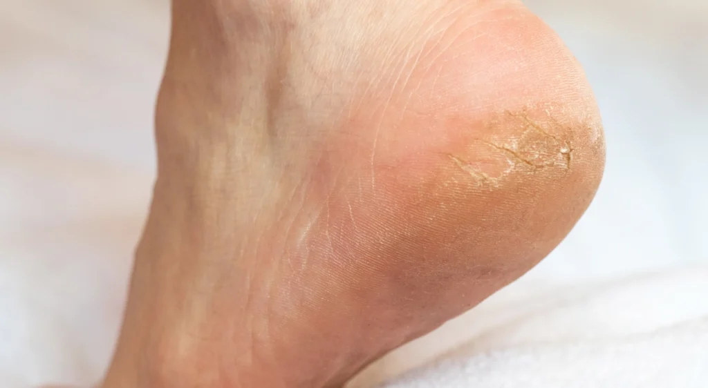
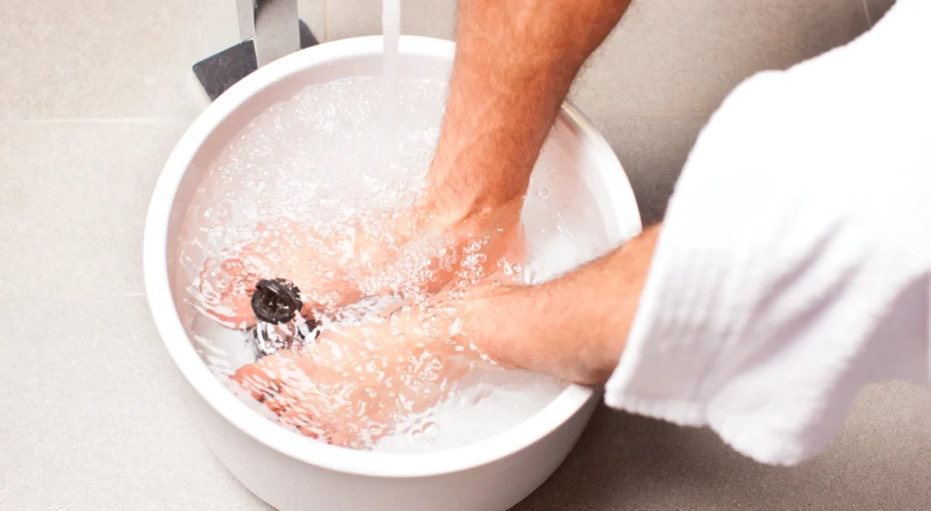
Hardness and Callus Removal
Calluses and corns on the feet are common problems that affect the quality of life…

Is it advisable to wear second-hand shoes?
The rise of the circular economy and sustainable practices has boosted interest in reusing products,…
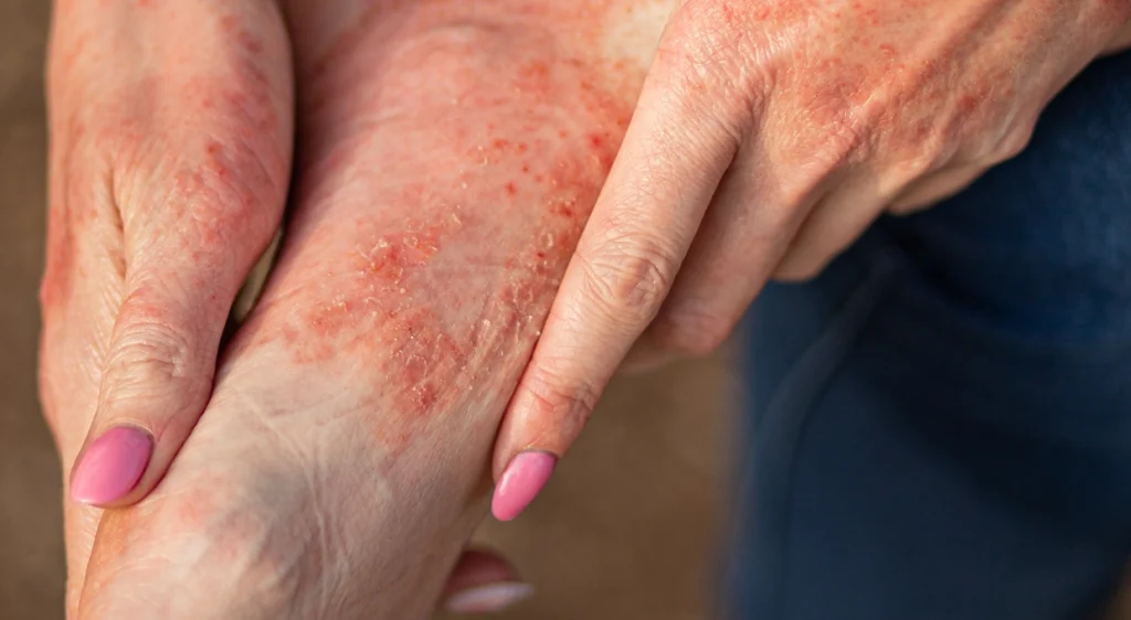
Eczema and Psoriasis on Feet
Eczema and psoriasis on the feet are two common dermatological conditions that, although they share…

Are podiatrists doctors in Spain? Everything you need to know about their training and competencies.
It is common for many people to wonder: are podiatrists doctors in Spain? The doubt…
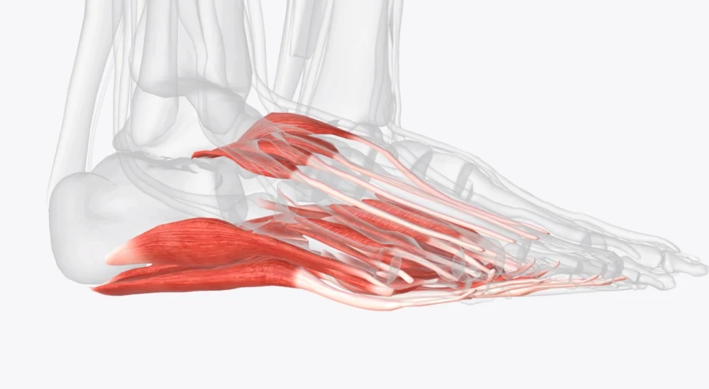
What muscles are in the foot? A complete guide to understand their function and care
The foot is a biomechanical marvel. It supports the entire weight of the body, adapts…
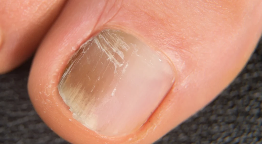
Brown toenail: causes, diagnosis and podiatric treatment
Brown nails is a clinical condition that is of concern to many patients, especially when…
How to maintain good foot hygiene at home
Foot hygiene is a fundamental pillar in maintaining foot health, preventing infections, avoiding discomfort and…

10 signs that you need medical attention for your feet
The feet support our body day after day, but we rarely give them the medical…