
SURGERY MY – SUBUNGUAL OSTEOCHONDROMA – ALICANTE
Subungual osteochondroma treatment: MIS surgery that preserves your nail
Percutaneous excision of the benign bone tumor under the nail – Preservation of the nail matrix in most cases – Same day walking under local anesthesia.
Backed by 45 years of experience in MIS foot surgery
Clinic founded in 1979
Three generations treating nail pathology and foot tumors in Alicante.
Certification MIS23BE03
Accredited by the American Board of Multispecialty Podiatry for minimally invasive surgery.
Multilingual service
We attend you in Spanish, English, German, French and Dutch.
What is subungual osteochondroma?
Subungual osteochondroma is a benign bone tumor that grows under the nail, almost always on the big toe or second toe. Although its name is frightening, it is not a cancer: it is a cartilaginous-osseous proliferation covered by a hyaline cartilage cap, perfectly delimited and of absolutely benign behavior. What makes it problematic is not its nature, but its location: developing between the distal phalanx and the nail bed, it lifts the nail, presses on nerve structures rich in nerve endings and causes pain disproportionate to the size of the lesion.
Osteochondroma represents the most frequent benign bone tumor of the skeleton, accounting for 35% to 40% of all tumors according to classical series (Murphey, RadioGraphics 2000). In its subungual variant it typically appears between the second and fourth decade of life, with a slight predominance in young active women. Several studies indicate that its location on the foot is a minority compared to femur or tibia, but, when it occurs, it is practically exclusive to the first toe.
At Clínica San Román we approach it with minimally invasive surgery: a millimetric incision allows access to the bone, milling the tumor and preserving in most cases the nail matrix, which is the structure responsible for the normal growth of the nail. This detail – preserving the nail – is the essential difference between our technique and classic open surgery.
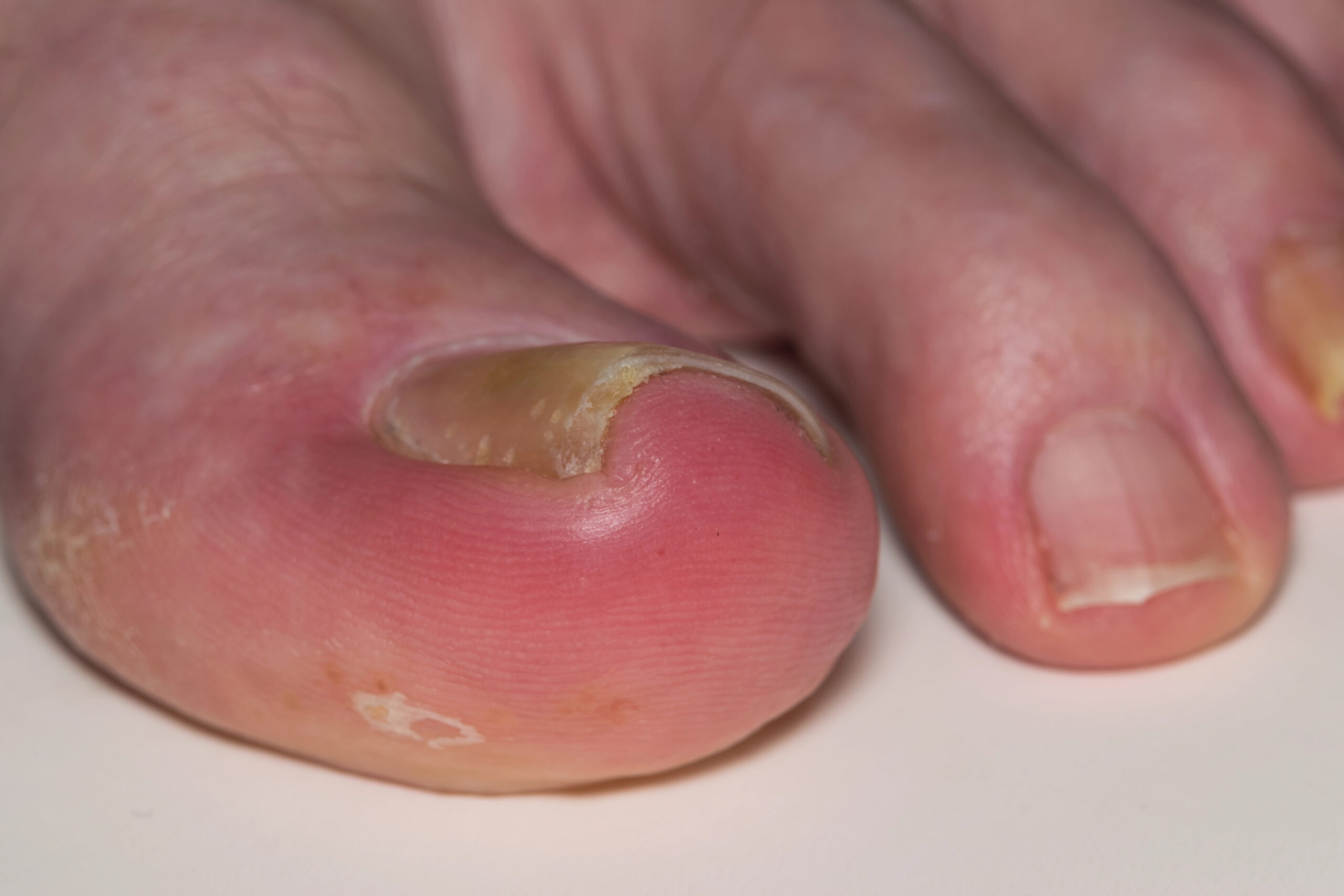
Symptoms: how to recognize a subungual osteochondroma?
The clinical picture is very characteristic once it is known. It usually starts as a small discomfort when wearing closed shoes or when resting the toe, which progresses for months until it becomes a stabbing pain with any friction. Usually the patient has previously consulted his family doctor or dermatologist believing that it is a wart, a mycosis or an ingrown toenail, and no previous treatment has helped.
- Selective pain when resting the toe or when wearing closed footwear, especially narrow toe shoes.
- Progressive lifting of the nail (distal onycholysis) with the appearance of “nail that does not stick”.
- Palpable hard lump under or lateral to the nail, with unmistakable bony consistency.
- Erythema, chronic inflammation and even ulceration of the nail bed due to continuous pressure.
- Pain on lateral squeezing of the finger, disproportionate to the visible size of the lesion.
- Nail deformity: curved, thickened or longitudinally grooved nail.
- Occasional bleeding or serous oozing due to microtrauma.
- In adolescents: pain that limits sports practice and makes it necessary to change footwear.
Why does subungual osteochondroma appear?
There is no single cause: we are talking about aberrant bone growth in which individual predisposition, repeated microtrauma and hormonal growth factors combine. Knowing these factors helps to understand why it recurs or why some patients develop it in apparently healthy feet.
🧬 Genetic predisposition
A small proportion of osteochondromas are part of hereditary multiple osteochondromatosis (multiple exostoses), an autosomal dominant disease associated with mutations in the EXT1 and EXT2 genes. When similar lesions appear in other bones of the skeleton or there is a family history, it is advisable to have a regular study. The isolated subungual form, on the other hand, is usually sporadic and does not involve familial risk.
👟 Repeated microtraumas
Narrow toe shoes, impact sports (running, soccer, dancing), practices such as ballet on tiptoes or climbing with climbing shoes compress the distal phalanx against the nail. These repeated microtrauma can activate foci of aberrant bone growth. This is why we see the picture especially in dancers, runners and women who wear narrow heels on a regular basis.
🌱 Aberrant bone growth
Osteochondroma is caused by an abnormal migration of growth cartilage that proliferates outside its usual place. This is why most cases debut in adolescence or young adulthood, when the growth plates are still active. Once the physis has closed, the tumor usually stops growing, although it may continue to give mechanical symptoms simply by its presence.
Diagnosis: how do we confirm subungual osteochondroma?
The diagnosis is clinical and radiological. The key factor is to confirm the bony nature of the lesion, rule out other subungual tumors and plan the surgery with millimetric precision. At Clínica San Román we protocolize the evaluation in three levels:
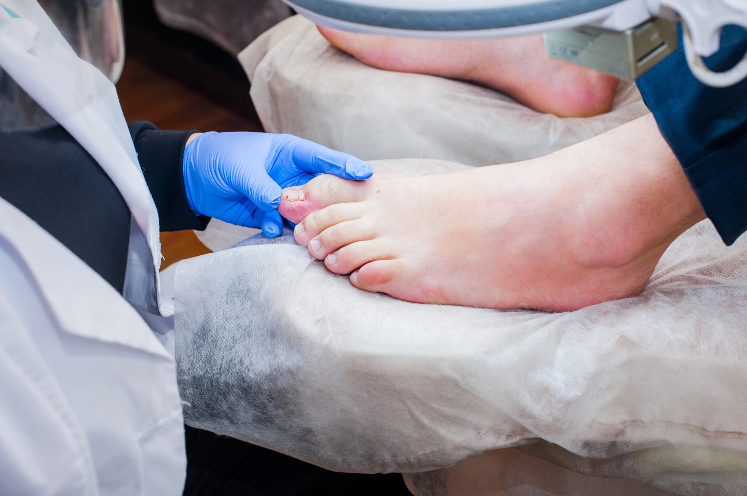
🔍 Clinical examination and dermoscopy
We inspect the nail, palpate the consistency of the bump and reproduce the pain with selective pressure. Dermoscopy helps to rule out pigmented lesions (subungual melanoma) and vascular pathology of the nail bed. We collect sports history, usual footwear and possible similar lesions in other fingers or limbs.
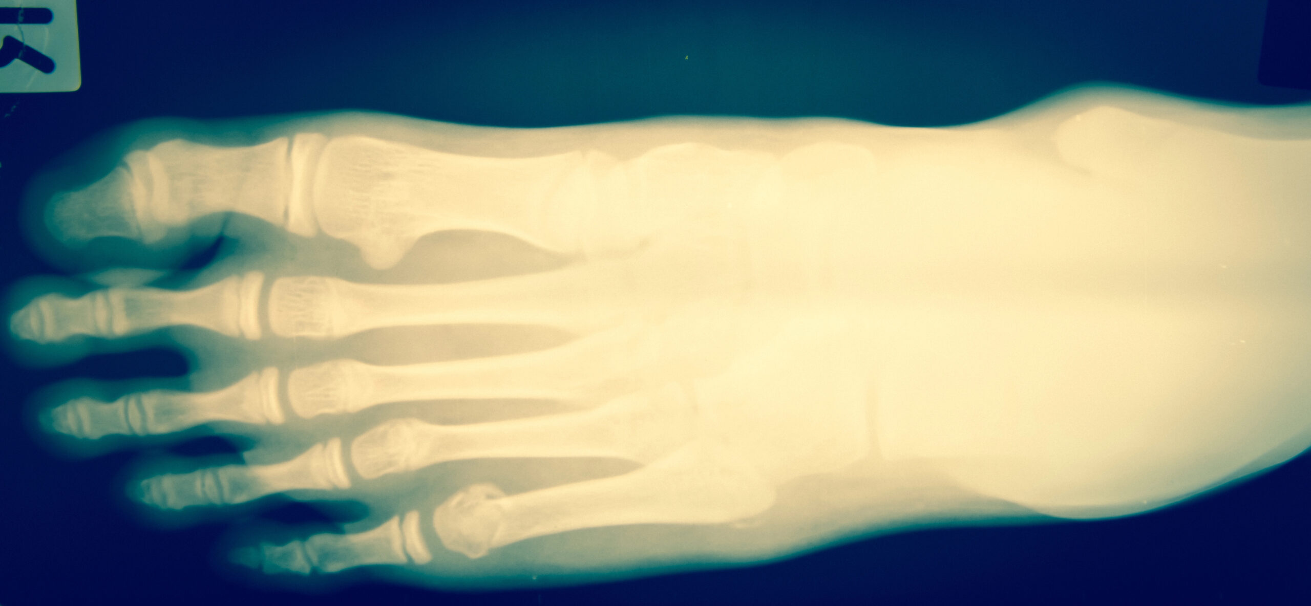
📷 Simple X-ray of the finger
Radiography is the pathognomonic test. We performed a dorsoplantar and lateral projection of the affected finger: the osteochondroma appears as a bony outgrowth with cortico-medullary continuity with the distal phalanx, covered by a cartilaginous cap. This detail – the medullary continuity – is what distinguishes it radiologically from subungual exostosis.
🔊 Ultrasound and magnetic resonance imaging
We reserve soft tissue ultrasound and MRI for cases of diagnostic doubt, suspicion of associated soft tumors or evaluation of the cartilaginous cap. MRI is also useful when symptoms are inconsistent with radiographic findings.
Treatment of subungual osteochondroma: stepwise protocol.
We apply a philosophy of less is more: we only escalate to the next step when the previous step does not resolve the symptomatology or the tests confirm clear surgical criteria. The good news is that, when surgery is required, MIS surgery resolves most cases in a single outpatient procedure.
1 Observation and handling of footwear
In asymptomatic lesions, casual findings or cases in which the patient does not want to undergo surgery, we propose a clinical-radiological follow-up every six months or yearly. We verify that the tumor does not grow, that it does not cause ulceration and that no signs of transformation appear. It is the option for small and well tolerated osteochondromas, especially in patients with surgical risk factors or in children whose growth cartilage is still active.
2 Shoe fitting and nail pads
When the pain is mild or moderate and there is a temporary contraindication to surgery (pregnancy, athlete in full competition season), we work on the mechanical factor: wide toe shoes with rigid soles, individualized silicone protectors that distribute the pressure and prevent direct contact of the tumor with the shoe, orthonyxia to correct associated curved nails and local cures of the bed if there is maceration. It is not a definitive solution, but it allows to gain time with quality of life.
3 MIS surgery – percutaneous excision preserving matrix
This is the definitive treatment and the one that most symptomatic patients undergo. The procedure lasts between 20 and 35 minutes, is performed under local anesthesia of the finger trunk, without prolonged tourniquet, and by a millimetric incision that in most cases respects the matrix and the nail plate. With specific MIS surgery instruments (chisels, drills and percutaneous files) we remove the tumor from its base, regularize the cortical part of the phalanx and send the piece to anatomopathological analysis for histological confirmation. The patient walks the same day with a functional bandage and post-surgical shoe.
Comparison: MIS surgery vs. traditional open surgery
| Criteria | Observation | Classic open surgery | MIS surgery (Clínica San Román) |
|---|---|---|---|
| Indication | Stable, asymptomatic osteochondroma | Large tumors, recurrent or with diagnostic doubt. | Majority of confirmed symptomatic osteochondromas |
| Invasiveness | Null | Partial or total avulsion of the nail + wide incision | Millimeter incision (3-4 mm), matrix preserved |
| Anesthesia | – | Local with sedation or regional | Local truncal toe |
| Time / sessions | Biannual check-ups | 30-60 min – outpatient or 24 h admission | 20-35 min – outpatient, same day discharge |
| Efficacy (pain resolution) | Not applicable | High (≈90 %) – recurrence 5-10 % in classic series | High – low recurrence with complete base resection |
Source: Multhopp-Stephens H and Walling AK, Orthopedics 1995; DaCambra MP et al, Foot Ankle Spec 2014; Murphey MD et al, RadioGraphics 2000. Individual results may vary.
Subungual exostosis vs. subungual osteochondroma
These are the two most common subungual bone lesions and patients (and many professionals) confuse them. The correct diagnosis changes the prognosis, the surgical technique and the risk of recurrence. This table summarizes the differences that we assess in consultation:
| Feature | Subungual exostosis | Subungual osteochondroma |
|---|---|---|
| Nature | Reactive bone reaction (not true tumor). Normal trabecular bone covered with fibrocartilage. | True benign bone tumor. Cancellous bone covered with hyaline cartilage cap. |
| Typical age | Adolescents and young adults (15-30 years) | Adolescents and young adults (10-35 years); associated with osteochondromatosis |
| Preferred location | Dorsomedial aspect of the first toe | Distal phalanx of the first toe (dorsal or lateral) |
| Cortical-medullary continuity | No (cortex does not continue with the bone of origin) | Yes (key radiological sign: cortex and medulla continue with the phalanx) |
| Cartilaginous cap | absent or very thin, fibrocartilage | Present, hyaline cartilage. Visible on MRI. |
| Traumatic history | Frequent (repeated microtraumatism) | Occasional. Bone growth factor predominates |
| Association with hereditary syndromes | No | Yes (hereditary multiple osteochondromatosis, EXT1/EXT2) |
| Transformation risk | Exceptional | Exceptional in subungual location; vigilance in large lesions |
| Recurrence after complete surgery | 5-10 % if the entire base is not resected | Very low with base and cartilage cap resection |
| Common technique at Clínica San Román | Percutaneous MIS excision | Percutaneous MIS excision with preservation of nail matrix |
Source: Murphey MD et al, RadioGraphics 2000; DaCambra MP et al, Clin Orthop Relat Res 2014. Individual results may vary.
Advantages of MIS subungual osteochondroma surgery
You walk the same day
Immediate discharge with post-surgical shoe with rigid sole. No plaster cast, no crutches required.
Local anesthesia
Truncal finger block. No general sedation, no prolonged fasting, no hospital admission.
Millimeter incision
3-4 mm incision that heals almost imperceptibly. Aesthetic result far superior to open surgery.
Preserves the nail
In most cases we respect the nail matrix. The nail is maintained or grows back normally.
High efficiency and low recurrence
Resolution of pain in the vast majority of patients. Low recurrence rate when the entire base is resected (Multhopp-Stephens and Walling, 1995).
Histological confirmation
All extracted specimens are sent to pathological anatomy. We confirm the diagnosis and rule out other lesions.
Have you been suffering for months from a painful lump under the nail that no treatment relieves?
Let us confirm exactly what it is. An X-ray and an assessment are enough to know if we are dealing with a subungual osteochondroma and to propose the treatment that will preserve your nail.
Request your free evaluationFrequently asked questions about subungual osteochondroma
We solve the most common doubts of our patients.
🔬 About the pathology.
Is it the same as a subungual exostosis?
No, although they are often confused. Subungual exostosis is a reactive bone reaction, almost always after microtrauma, without true cartilaginous cap. Osteochondroma is a true benign bone tumor, with hyaline cartilage and cortico-medullary continuity with the phalanx. The cause, radiological image, typical age and risk of recurrence change.
Can osteochondroma be malignant?
Osteochondroma is, by definition, benign. There is a very rare malignant transformation to chondrosarcoma (less than 1% in solitary forms and higher in multiple osteochondromatosis), but it has practically never been described in subungual location due to its small size. Even so, at Clínica San Román we always send the extracted piece to pathological anatomy for histological confirmation.
Why does it hurt so much if it is benign?
Pain does not depend on the size or nature of the tumor, but on its location. The nail bed is an area rich in nerve endings and vessels. A bony excrescence of a few millimeters lifts the nail, compresses the receptors and creates a chronic inflammation of the bed.
When is it usually diagnosed?
The peak of presentation is between 15 and 35 years of age, coinciding with still active growth cartilages and with the most intense sports or work practice. Several studies indicate a slight female predominance. In adolescent athletes it usually takes months to be diagnosed because it is initially confused with ingrown toenail or sporting injury.
🏥 Treatment
Must the nail always be removed?
No. The great advantage of MIS surgery is that we preserve the nail in most cases. We access the tumor through a lateral or dorsal millimeter incision that respects the nail matrix, we mill the bone and the nail remains intact.
How long does the surgery last?
The operation itself lasts between 20 and 35 minutes. Adding preparation, truncal local anesthesia of the toe and postoperative functional bandage, the total time in the operating room is about 60 minutes. It is ambulatory: you go in and out on your foot the same day.
Can I put my foot down immediately?
Yes, you walk the same day with a stiff-soled post-surgical shoe, which distributes the load and protects the toe for the first 2-3 weeks. Impact sports are typically resumed between the 4th and 6th week.
🏃 Recovery and prevention
Does the nail grow back normally?
In most cases yes, because we preserve the matrix, which is the “factory” of the nail. If there was previous nail dystrophy due to chronic pressure from the tumor, the nail usually recovers its shape as the tissue reorganizes itself, within 6 to 12 months.
Can it recur after surgery?
The risk of recurrence is low when the base of the tumor and its cartilaginous cap are completely resected. The classic literature (Multhopp-Stephens and Walling, 1995) reports recurrence rates of less than 5-10% with adequate techniques. We recommend revisions at 6 and 12 months with control radiography.
Can I prevent its occurrence?
There is no specific prevention of osteochondroma because its origin is constitutional. We can minimize the microtraumatisms that act as a trigger: wide toe shoes and breathable materials, shock absorbing soles in impact sports, correct nail trimming and regular podiatric check-ups if you practice ballet, athletics, soccer or climbing.
Scientific references
- Murphey MD, Choi JJ, Kransdorf MJ, Flemming DJ, Gannon FH. Imaging of osteochondroma: variants and complications with radiologic-pathologic correlation. RadioGraphics. 2000;20(5):1407-34. [PMID: 10992031]
- Multhopp-Stephens H, Walling AK. Subungual exostosis. Orthopedics. 1995;18(4):385-7. [PMID: 7596246]
- DaCambra MP, Gupta SK, Ferri-de-Barros F. Subungual exostosis of the toes: a systematic review. Clin Orthop Relat Res. 2014;472(4):1251-9. [PMID: 24249538]
- Carroll RE, Chance JT, Inan Y. Subungual exostosis in the hand. J Hand Surg Br. 1992;17(5):569-74. [PMID: 1479252]
- Bovée JVMG. Multiple osteochondromas. Orphanet J Rare Dis. 2008;3:3. [PMID: 18271966]
- de Palma L, Gigante A, Specchia N. Subungual exostosis of the foot. Foot Ankle Int. 1996;17(12):758-63. [PMID: 8973898].
Information prepared by the medical team of Clínica San Román. Center No. 5357 Autonomous Registry of Health Centers, Services and Establishments of the Valencian Community. This page is for information purposes only and does not replace the individual clinical assessment.
Don’t let osteochondroma pain control your day-to-day life
An X-ray and an evaluation are enough to confirm the diagnosis and plan the MIS surgery that preserves your nail. We have been doing it for 45 years in Alicante.
- 📞 +34 965 921 156
- ✉️ info@clinicasanroman.com
- Av. del Dr. Ramón y Cajal 1, 03001 Alicante