ADVANCED PODIATRY · HEEL SPUR · ALICANTE
Calcaneal spur: effective pain-free treatment
Custom insoles. Shockwave therapy. Percutaneous surgery if necessary. Walk the same day.
If you feel a sharp pain in your heel every morning when you take your first steps—if you’ve been experiencing that sensation of stepping on a nail for months and your walking has been getting worse—you likely have a heel spur or chronic plantar fasciitis. At Clínica San Román, we follow a step-by-step protocol: first, conservative treatment (insoles + shockwave therapy), and only if that fails, percutaneous surgery under local anesthesia. 90% of our patients improve without needing surgery.
⭐ 4.8/5 (190 reviews) · 🏅 Cert. MIS23BE03 · 🇪🇺 5 languages
Backed by 45 years of experience
45+ years old
Family clinic founded in 1979
4.8 ★
190 reviews on Google
MIS 🏅
First in Europe to offer an MIS subspecialty (Cert. MIS23BE03)
🇪🇺 5
Languages: ES · EN · DE · FR · NL
What is a heel spur?
A heel spur is an abnormal bony growth (osteophyte) that forms on the underside of the heel bone (calcaneus), in the area where the plantar fascia attaches. It is like a small bony spur that grows horizontally toward the toes and can measure between 1 and 20 millimeters.
The most important thing to know is that a heel spur itself does not always cause pain. In fact, X-rays show that many people have heel spurs without experiencing any symptoms. In most cases, the pain is caused by associated plantar fasciitis: a chronic inflammation of the plantar fascia that accompanies the heel spur.
It is estimated that heel spurs affect 10% of the population at some point in their lives, with the condition being most common among people aged 40 to 60. Approximately 89% of patients with chronic plantar fasciitis have a heel spur detectable on X-ray. It is one of the most common causes of plantar talalgia (heel pain) that we treat in our practice.
Symptoms: How do I know if I have a heel spur?
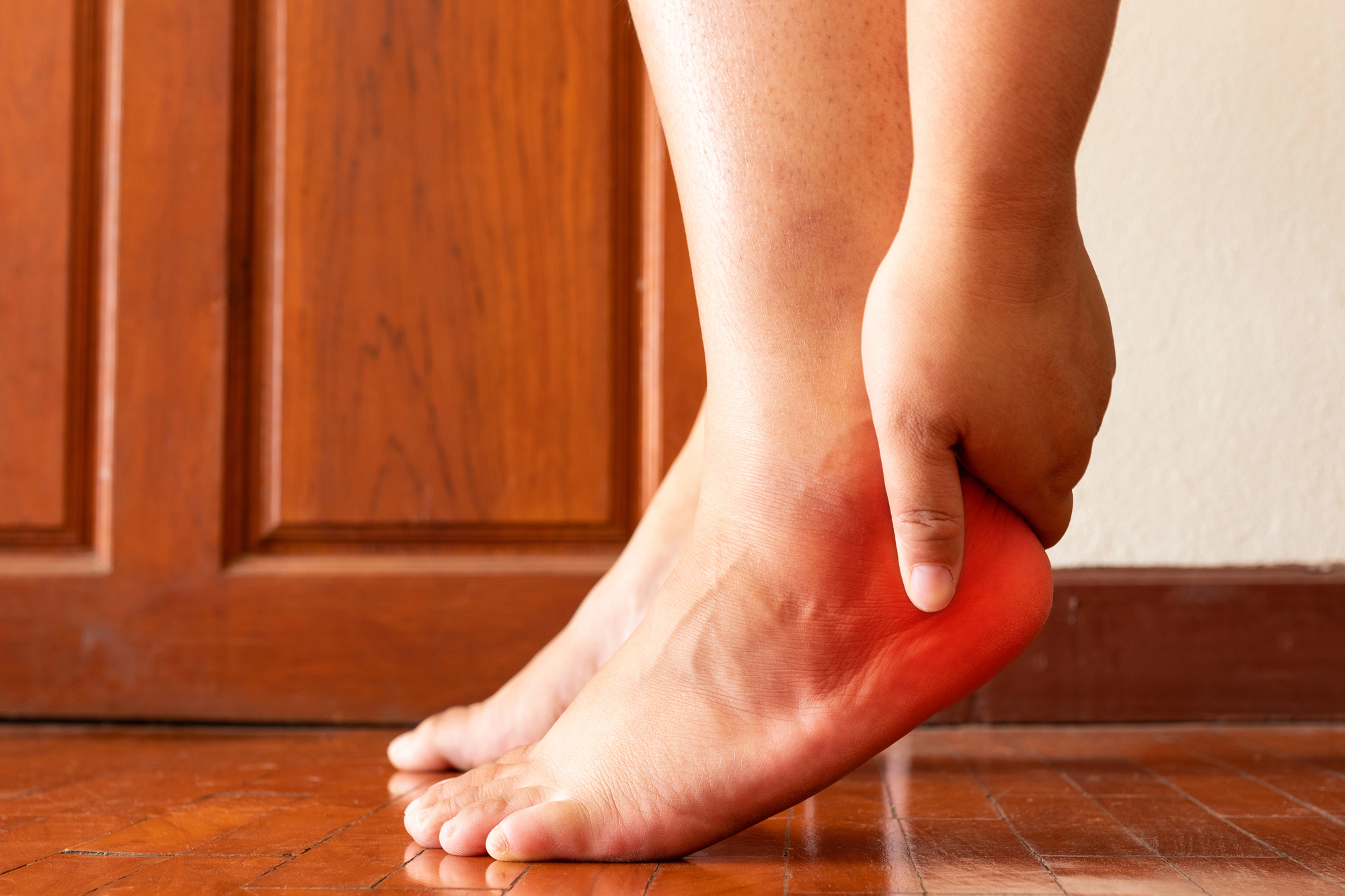
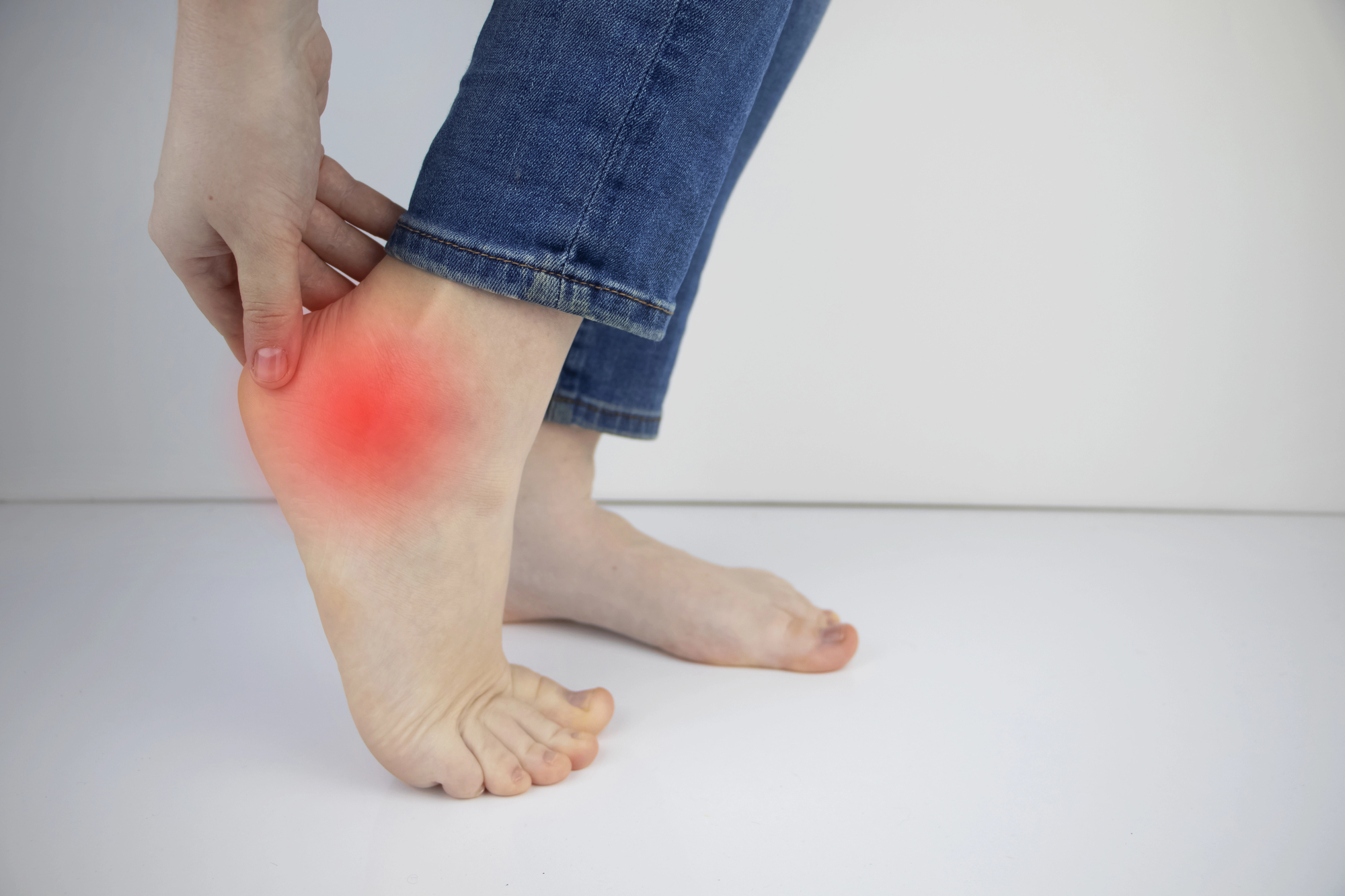
The most characteristic symptom is what is known as “morning heel pain”: a sharp, intense pain in the heel when getting out of bed in the morning or after sitting for a long time. The pain usually improves after walking for a few minutes (when the fascia “warms up”), but it returns at the end of the day or after prolonged activity.
- A sharp pain in the center or inner edge of the heel —as if you’d stepped on a nail or a rock when taking your first steps
- Pain that worsens after rest —when getting up, after sitting, or when getting out of the car (a typical pattern of plantar fasciitis)
- Pressure sensitivity — pain when pressure is applied to the inner part of the heel (medial calcaneal tuberosity)
- Morning stiffness — a feeling of tightness in the sole of the foot upon waking that gradually eases with movement
- Compensatory limping — when trying to avoid putting weight on the heel, the gait is altered and secondary pain may develop in the knee, hip, or back
⚠️ Important: Not all heel pain is caused by a heel spur. Other conditions such as calcaneal stress fracture, fat pad atrophy, Baxter’s nerve compression, or tarsal tunnel syndrome can cause similar symptoms. An accurate diagnosis through clinical examination and imaging is essential.
Why does a heel spur form?
A heel spur forms as the bone’s response to chronic mechanical stress. When the plantar fascia repeatedly pulls on its attachment to the heel bone (due to overuse, poor biomechanics, or both), the body deposits calcium in the stressed area and forms a bony protrusion. It is a slow process that can take months or years.
🏋️ Biomechanical factors
- Flatfoot or excessive pronation — increases the strain on the plantar fascia with every step
- Cavus foot — an excessively high arch that concentrates pressure on the heel and forefoot
- Shortening of the Achilles tendon — limits ankle dorsiflexion and puts strain on the plantar fascia
- Leg length discrepancy — one leg being longer than the other causes asymmetric stress
⚙️ Lifestyle factors
- Overweight or obesity — excess weight increases the strain on the heel with every step (a BMI of > 30 is a major risk factor)
- Prolonged standing — occupations that require standing for more than 8 hours a day (hospitality, healthcare, retail)
- Inappropriate footwear —flat soles, no cushioning, or heavily worn shoes
- High-impact sports — road running, jumping, and quick changes of direction on hard surfaces
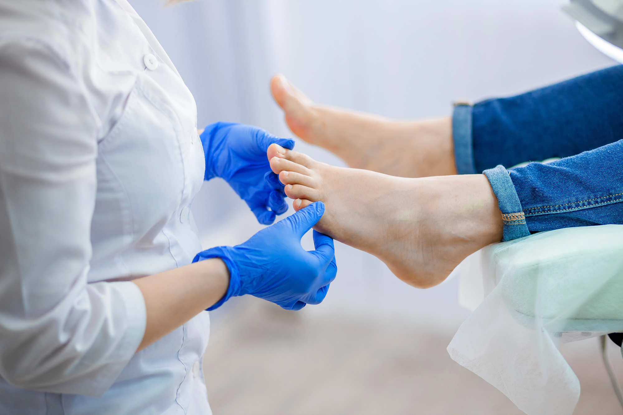
Diagnosis: We’ll determine the exact cause of your pain
An accurate differential diagnosis is essential, because not all heel pain is caused by a heel spur. At Clínica San Román, we perform:
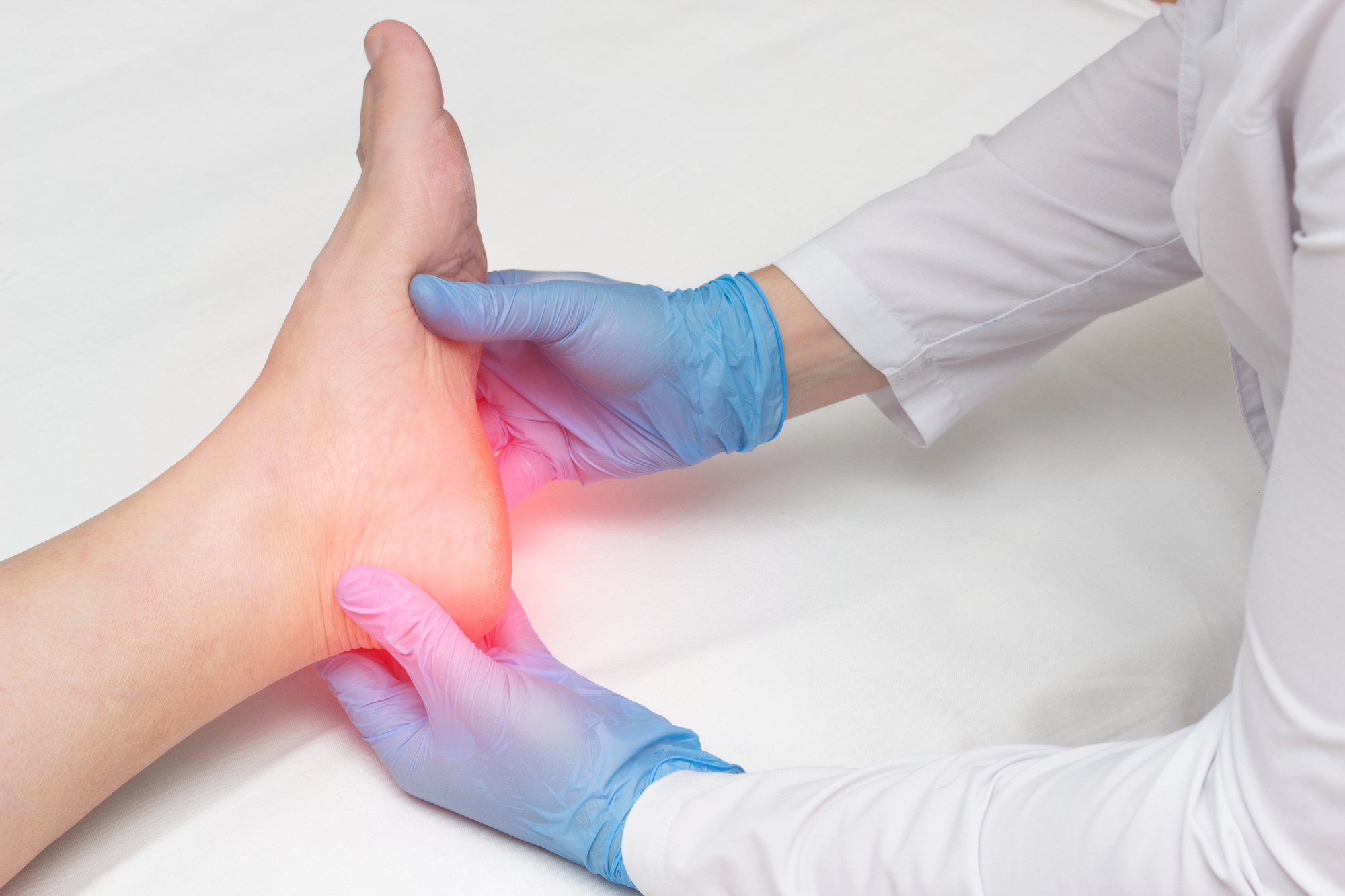
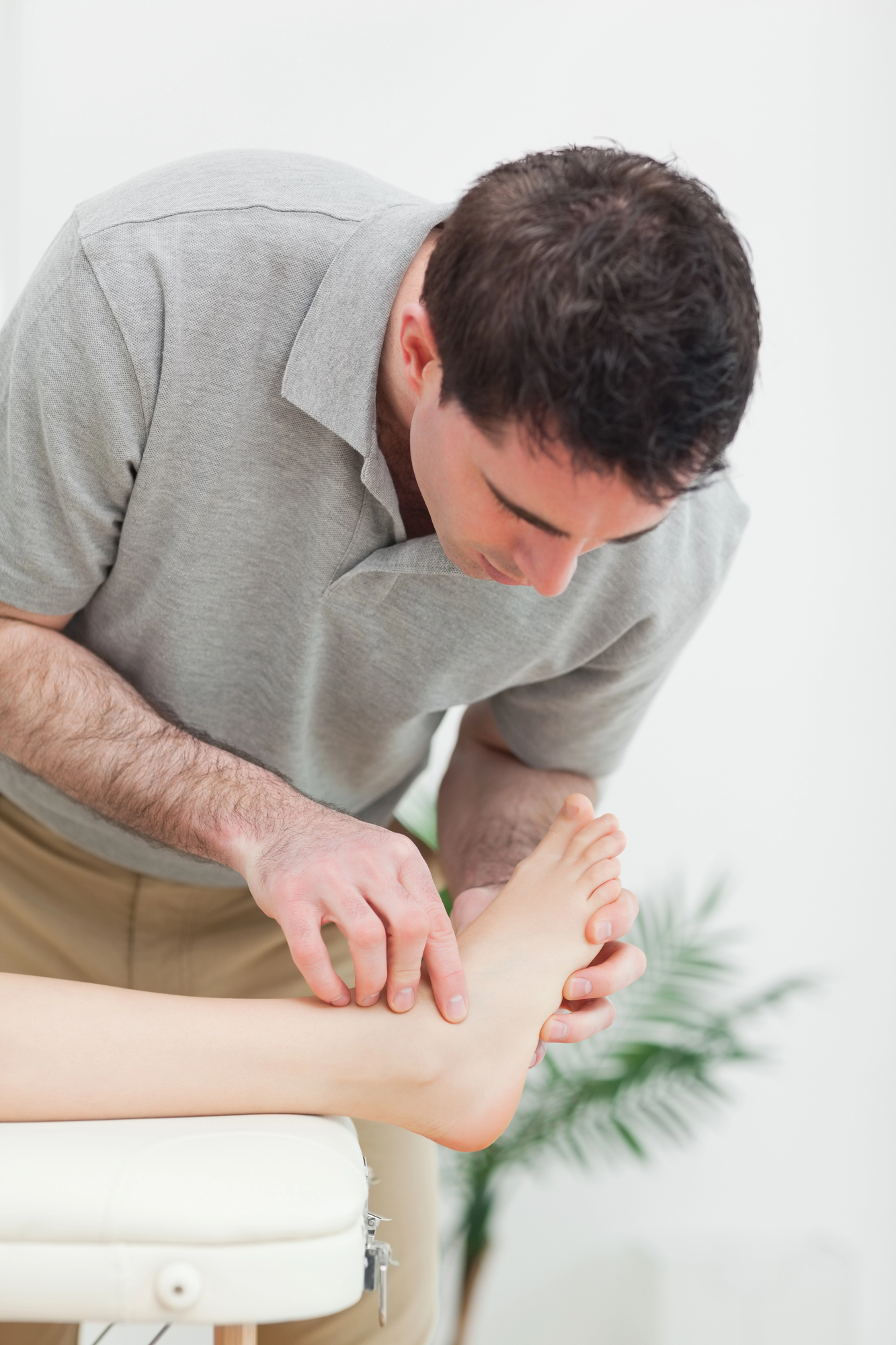
🔍 Clinical examination
Palpation of the medial calcaneal tuberosity, Windlass test (dorsiflexion of the big toe), assessment of ankle range of motion, and biomechanical gait analysis. Selective pressure on the medial insertion of the fascia reproduces the typical pain.
📷 Lateral X-ray
It allows for visualization of the heel spur, measurement of its size, and classification of its type (Type A—above the fascia; Type B—within the fascia). It also rules out stress fractures of the calcaneus or other bone conditions.
🔊 Soft tissue ultrasound
We measure the thickness of the plantar fascia (normal: 4 mm). It detects thickening, partial tears, retrocalcaneal bursitis, and inflammatory fluid. It is a quick, painless, and radiation-free test.
Treatment of heel spurs: a stepwise protocol
Our approach follows the clinical practice guidelines of the APTA (American Physical Therapy Association, 2023 update): a stepwise protocol that begins with conservative treatment and reserves surgery for refractory cases.
Step 1 — Conservative treatment (0–3 months)
- Custom orthotic insoles with heel cups, retrocalcaneal bars, and arch supports—manufactured at our clinic following a biomechanical assessment
- Specific stretches for the plantar fascia and the soleus-gastrocnemius complex (guided home exercise program)
- Appropriate footwear with heel cushioning and a semi-rigid sole
- Manual therapy and ankle joint mobilization if there is a lack of dorsiflexion
Step 2 — Advanced therapies (3–6 months)
- Extracorporeal shock wave therapy (ESWT) — stimulates tissue regeneration in the plantar fascia. Treatment protocol consisting of 3–5 sessions. Proven efficacy comparable to surgery in appropriate cases
- Ultrasound-guided corticosteroid injection (in selected cases, up to 2–3 injections) to control acute inflammation
Step 3 — Percutaneous surgery (if the previous step fails)
- Percutaneous partial plantar fasciotomy — release of the plantar fascia at its insertion using minimally invasive surgical (MIS) instruments through a millimeter-long incision
- Osteophyte removal — resection of the osteophyte using a surgical drill under fluoroscopic guidance
- Calcaneal decompression — microperforations that stimulate bone revascularization
- Local anesthesia — no risks associated with general anesthesia. The patient is able to walk the same day wearing postoperative footwear
Treatment Options: A Comparison
The choice of treatment depends on the severity, duration of symptoms, and response to previous measures.
| Feature | Exercises + Stretches | Shock Waves (ESWT) | Echoguided infiltration | Minimally invasive percutaneous surgery |
|---|---|---|---|---|
| Indication | First row (all) | Conservative treatment failure ≥ 3 months | Severe acute pain | Complete failure ≥ 6–12 months |
| Invasiveness | None | Non-invasive | Minimally invasive | Percutaneous (millimeter-sized) |
| Anesthesia | Not applicable | Not applicable | Local (period) | Local (heel) |
| Sessions / Time | Continuous use | 3–5 sessions (once a week) | 1–3 injections | 1 session (30 min) |
| Load after treatment | Immediate | Immediate | Immediate | Immediate (postqx footwear) |
| Back to Business | Immediate | Immediate | 24–48 hours | 4-6 weeks |
| Estimated effectiveness | 70-80% | 65-80% | Temporary (3–6 months) | 85-95% |
| Side effects | None | Temporary pain | Risk of fascia rupture | Minimums |
* The efficacy rates are based on published evidence (APTA 2023, Jiang et al. 2024). Individual results may vary.
Benefits of Percutaneous Heel Spur Surgery
When surgery is necessary, our minimally invasive surgery offers clear advantages over traditional open surgery:
🚶
You walk the same day
Immediate ambulation in postoperative shoes upon discharge from the clinic
💉
Local anesthesia
No risks associated with general anesthesia, no hospitalization, and no need to fast beforehand
🔬
Millimeter incision
Minimal tissue trauma, lower risk of infection, and an imperceptible scar
⏱️
Rapid recovery
Regular shoes after 3 weeks. Sports activities after 6–8 weeks
🎊
Highly effective
85–95% success rate. Significant improvement on the AOFAS and VAS pain scales
🔄
Triple action
Fasciotomy + removal of the bone spur + decompression in a single procedure
Prevention: How to Prevent Heel Spurs from Developing or Recurring
If you have already had a heel spur or have risk factors, these measures can help prevent its onset or recurrence:
👟 Suitable footwear
Wear shoes with good heel cushioning, a semi-rigid sole, and a sturdy heel counter. Avoid flat shoes (ballet flats, flip-flops) and high heels. Replace your running shoes every 600–800 km or every 6 months.
🧘 Daily Stretches
Stretch your plantar fascia and Achilles tendon every morning before getting out of bed and after each workout. Do 3 sets of 30 seconds per stretch. Research shows that stretching can reduce the recurrence of pain by up to 52%.
⚖️ Weight management
Maintain a healthy body weight. Every extra pound increases the pressure on your heel when you walk. Weight loss, when recommended, is one of the most effective ways to prevent plantar fasciitis and heel spurs.
Have you been dealing with heel pain for months? We can help.
Heel spurs are a condition with an excellent prognosis when diagnosed and treated properly. However, many patients wait months—or even years—before seeking help, and during that time, the pain affects their gait, quality of life, and mood.
At Clínica San Román, Dr. Israel San Román and Dr. José Manuel San Román offer you a free evaluation with radiological diagnosis to determine the exact cause of your pain and recommend the best treatment. Our step-by-step protocol helps 90% of patients improve without the need for surgery.
📍 Downtown Alicante · 📞 +34 965 921 156 · 📧 info@clinicasanroman.com
Frequently Asked Questions About Heel Spurs
We solve the most common doubts of our patients. If your question isn’t listed here, please contact us and we’ll get back to you with no obligation.
📚 Scientific references (PubMed)
- Martin RL, Davenport TE, Reischl SF, et al. Heel Pain — Plantar Fasciitis: 2023 Update. J Orthop Sports Phys Ther. 2023;53(12):CPG1-CPG39. doi:10.2519/jospt.2023.0303. PubMed 38037331
- Jiang Y, Li H, Zhang Q, et al. Clinical Study of a Four-Step Program for the Treatment of Plantar Fasciitis with Bone Spurs. Orthop Surg. 2024;16(6):1456-1463. doi:10.1111/os.14061. PubMed 38693719
- Menz HB, Zammit GV, Landorf KB, Munteanu SE. Plantar calcaneal spurs in older adults: longitudinal traction or vertical compression? J Foot Ankle Res. 2008;1(1):7. doi:10.1186/1757-1146-1-7. PubMed 18822152
- Johal KS, Milner SA. Plantar fasciitis and the calcaneal spur: Fact or fiction? Foot Ankle Surg. 2012;18(1):39-41. doi:10.1016/j.fas.2011.03.003. PubMed 22326003
- Özdemir H, Söyüncü Y, Özgörgen M, Dabak K. Effects of changes in heel fat pad thickness and elasticity on heel pain. J Am Podiatr Med Assoc. 2004;94(1):47-52. doi:10.7547/0940047. PubMed 14729991
- Rompe JD, Furia J, Weil L, Maffulli N. Shock wave therapy for chronic plantar fasciopathy. Br Med Bull. 2007;81-82:183-208. doi:10.1093/bmb/ldm005. PubMed 17456546
- Aqil A, Siddiqui MR, Solan M, Redfern DJ, Gulati V, Cobb JP. Extracorporeal shock wave therapy is effective in treating chronic plantar fasciitis: a meta-analysis of RCTs. Clin Orthop Relat Res. 2013;471(11):3645-3652. doi:10.1007/s11999-013-3132-2. PubMed 23813184
- Kudo P, Dainty K, Clarfield M, Coughlin L, Lavoie P, Lebrun C. Randomized, placebo-controlled, double-blind clinical trial evaluating the treatment of plantar fasciitis with an extracorporeal shockwave therapy (ESWT) device. Foot Ankle Int. 2006;27(10):790-796. doi:10.1177/107110070602701006. PubMed 17054878
Don’t let heel pain take over your daily life
If you’ve been limping for months, if you’ve given up on walking, running, or enjoying a stroll— you don’t have to keep living like this. Heel spurs can be effectively treated, and in most cases, surgery isn’t even necessary.
San Román Clinic: Over 45 years of caring for your feet. Free consultation.