
Foot imaging study in Alicante
A good treatment begins with an accurate diagnosis. At Clínica San Román we have our own foot imaging technology -fluoroscopy, digital radiography and vascular EcoDoppler- which allows us to see what is happening inside, plan the surgery safely and make an accurate follow-up, all in the same center and without having to travel.
Proprietary technology
Fluoroscopy, digital radiography and EchoDoppler in the clinic.
Low radiation dose
Digital equipment with radiation safety control.
Results on DVD
We provide you with all your studies to keep them.
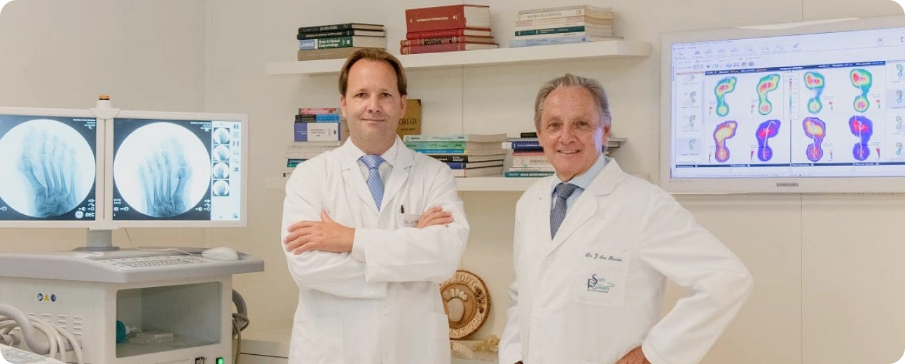
45+ years of experience
Diagnosis at the service of MIS foot surgery.
Our diagnostic technology
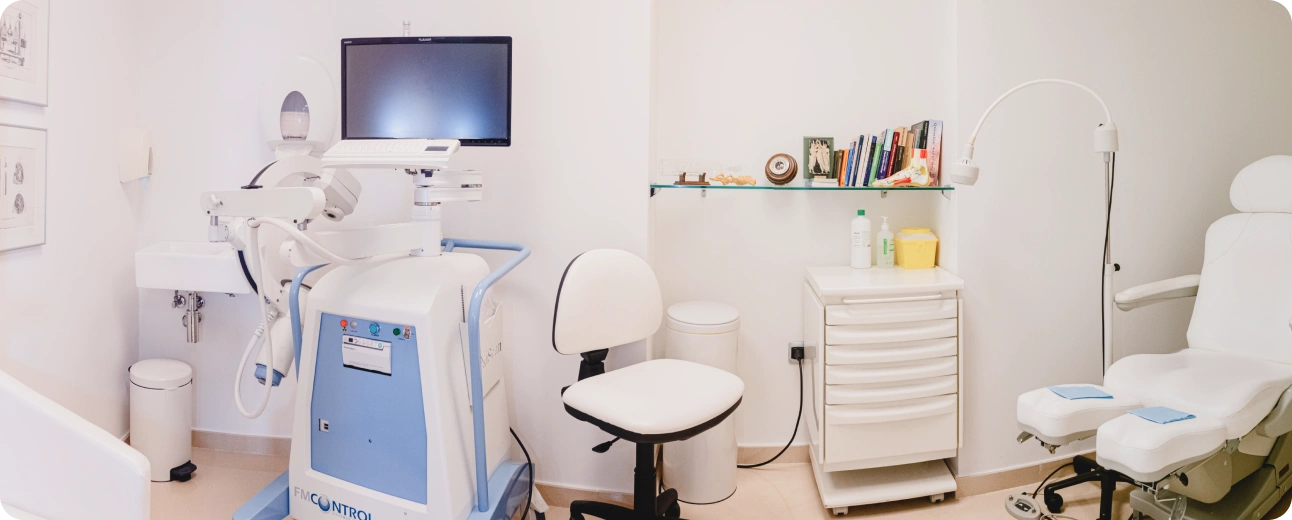
Fluoroscopy
Real-time radiology that guides percutaneous surgery and assesses post-surgical evolution.
Digital radiography
High-definition bone imaging in multiple projections, including loading radiography.
4D Vascular Doppler ultrasound
Study of blood flow and soft tissue for safer surgery.
Fluoroscopy analysis
Fluoroscopy is a radiodiagnostic technique that shows the bone structure of the foot in real time, with a low radiation dose. It is the key tool in minimally invasive surgery (percutaneous): it allows the surgeon to guide each gesture with precision through millimetric incisions. It is also very useful for post-surgical assessment. We have two XiScan fluoroscopes in the clinic.
Digital radiological study
We perform high definition digital radiography of the bone structure of the foot with Brivo equipment from General Electric, with minimum radiation dose. We obtain the necessary projections -standard, special and loaded-for a complete diagnosis. At the end, we deliver all your studies in a DVD from Clínica San Román.
Vascular EchoDoppler study
EchoDoppler is a hemodynamic study that analyzes blood flow and the state of the soft tissues of the foot using ultrasound, without radiation. It helps us to plan safer surgery and to accurately assess and treat lesions such as neuromas, cysts or guided infiltrations.
Radiation safety
Your safety is a priority. We work with low-dose digital equipment, which is reviewed annually and supervised by a Technical Unit of Radiological Protection (UTPR), complying with current safety regulations. We perform only the tests necessary for your diagnosis.
Why choose Clínica San Román
- Diagnosis and treatment in the same center: no waiting or travel to another place.
- Technology at the service of minimally invasive surgery, of which we are European pioneers.
- 45+ years of experience and average rating of 4.8/5 with over 191 reviews.
- Multilingual service (Spanish, English, German, French and Dutch).
Frequently asked questions about the imaging study
What foot imaging tests do you perform?
We have fluoroscopy (real-time radiology), high definition digital radiography and vascular EcoDoppler study. This allows an accurate diagnosis and planning of surgery with greater security, without having to travel to another center.
How much radiation do I receive?
Digital radiography and fluoroscopy are performed with low doses. Our equipment is checked annually and is supervised by a Technical Unit of Radiological Protection (UTPR), following the current safety regulations.
Do I get my test results?
Yes, we provide the patient with all his studies on a DVD from Clínica San Román, so that he has his documentation and can share it with other professionals if he wishes to do so.
What is fluoroscopy and what is it used for?
It is a real-time radiology that shows the bone structure of the foot in motion. It is key in minimally invasive surgery, because it allows to guide the intervention with precision, and also to assess the evolution after surgery.
What is the EchoDoppler study used for?
It studies blood flow and soft tissues of the foot. It helps to plan safer surgery and to assess and treat lesions such as neuromas, cysts or guided infiltrations.
Scientific support
Diagnostic imaging of the foot is well established in the literature. The loading radiography is basic to assess deformities such as hallux valgus and to plan surgery, and ultrasound reaches a high sensitivity (around 95% in expert hands) in the diagnosis of Morton’s neuroma, being of great help in the study of soft tissues.
Scientific references
- Imaging evaluation of hallux valgus. 2020. PMC: PMC7302897.
- Morton’s neuroma – current concepts review. 2020. PMC: PMC7211826.
- Radiological approach to metatarsalgia in current practice: an educational review. Insights into Imaging. 2025.
This page is for information purposes only and does not replace the individual clinical assessment. Each case requires a personalized examination.
Request your assessment with diagnostic imaging
If you need an accurate diagnosis of your foot or a second opinion, we have the technology to give it to you the same day. Call us at (+34) 965 921 156 or request an appointment.
