What is Percutaneous Surgery?
Percutaneous foot surgery, also known as minimally invasive surgery (MIS) or minimal incision surgery, represents a significant evolution in the surgical treatment of foot deformities. Through small incisions of just 2-5 millimeters, this technique makes it possible to correct various foot pathologies with considerably less surgical trauma than traditional techniques.
Unlike conventional open surgery, which requires large incisions of 5-10 centimeters to directly visualize the anatomical structures, the percutaneous technique works with specialized instruments and continuous radiological control. The surgeon uses a fluoroscope (real-time X-ray equipment) that allows visualization of the bone structures during the entire operation, guiding each cut and each movement of the instruments with millimetric precision.
This methodology allows the necessary bone corrections to be made while preserving the surrounding soft tissues as much as possible: muscles, tendons, ligaments and especially the vascularization of the foot. The preservation of the blood supply is one of the key factors that explains the generally faster recovery of this technique compared to traditional open approaches.
For what deformities is it indicated?
Percutaneous surgery has demonstrated efficacy in the treatment of multiple foot pathologies, although its main indication focuses on forefoot deformities of mild to moderate severity.
Hallux valgus or bunion is probably the most frequently treated deformity by percutaneous surgery. This technique is particularly appropriate when the intermetatarsal angle (IMA) is less than 15 degrees and the hallux valgus angle (HVA) does not exceed 40 degrees. In these cases, percutaneous osteotomies allow realignment of the first metatarsal and the big toe without the need to open the tissues extensively, respecting the joint capsule and minimizing damage to noble structures.
Hammertoes or claw toes also respond favorably to percutaneous techniques, especially when the deformity still retains some flexibility. By means of small tenotomies (tendon cuts) and osteotomies (bone cuts), normal finger alignment can be restored without the visible scars of open surgery.
Metatarsalgia, that intense pain under the metatarsal heads that hinders walking, can be corrected by means of shortening or elevation osteotomies of the metatarsal heads, thus redistributing the pressures during walking. These interventions are performed through incisions of 3-4 millimeters, practically imperceptible once healed.
Hallux rigidus, or osteoarthritis of the first toe, in its early stages may benefit from percutaneous cheilectomy. This procedure consists of removing the osteophytes (bony spikes) that limit joint movement, significantly improving range of motion and reducing pain.
In the midfoot and hindfoot, percutaneous surgery can treat exostoses (painful bony prominences), such as the so-called “tailor’s bunion” in the fifth metatarsal, calcaneal spurs resistant to conservative treatment by releasing the plantar fascia, and Haglund’s deformity (posterior heel prominence) by percutaneous resection of the prominent bone.
Proven Advantages of Percutaneous Surgery
The main advantage of percutaneous surgery lies in its minimally invasive nature. While traditional open surgery requires large incisions to allow direct visualization of anatomical structures, percutaneous techniques work through small portals of only 2-5 millimeters. This seemingly simple difference has profound implications in terms of surgical trauma, recovery and cosmetic results.
Soft tissue preservation is essential. During open surgery, it is necessary to section or retract muscles, tendons and ligaments to gain access to the bone. This process, although controlled, inevitably damages structures that must subsequently heal. In percutaneous surgery, specialized instruments make it possible to work between the tissues without sectioning them, respecting the original anatomy. This preservation translates into less postoperative pain, less inflammation and, crucially, less risk of scar adhesions that can limit future mobility.
Minimal scarring represents another objective benefit. The small incisions of 2-5 millimeters are virtually unnoticeable once healing is complete, especially when compared to the 5-10 centimeter linear scars characteristic of open surgery. For many patients, particularly those who wear open shoes or engage in activities where the feet are exposed, this aesthetic aspect is of significant importance.
Postoperative pain is usually more manageable in percutaneous surgery. Multiple comparative studies have documented that patients who undergo minimally invasive techniques report lower levels of pain during the first postoperative weeks, with less need for strong analgesics. Although discomfort exists and requires standard analgesic treatment, the intensity tends to be lower than in open approaches.
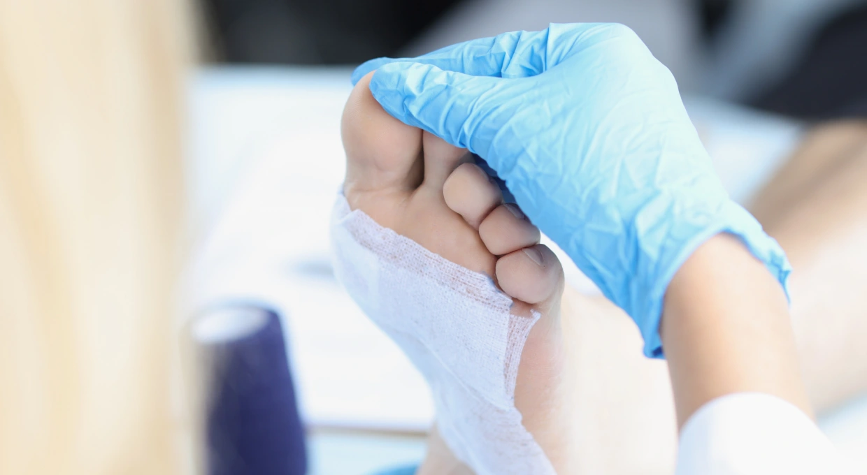
Functional recovery presents, in many cases, a more favorable evolution. The possibility of starting to ambulate from the first day with specialized post-surgical footwear, less inflammation of soft tissues and preservation of vascular structures contribute to an earlier return to daily activities. However, it is essential to clarify that “faster recovery” does not mean “immediate recovery”. Bone healing requires the same physiological time regardless of the technique used, typically 8-12 weeks for primary healing.
The outpatient nature of the procedure represents an organizational and economic advantage. Most percutaneous interventions are performed under local anesthesia and conscious sedation, without requiring general anesthesia. After an observation period of 1-2 hours in the recovery room, the patient can return home the same day. This feature not only reduces hospital costs, but also allows the patient to recover in a familiar environment, which is generally more comfortable than a hospital room.
Finally, the lower risk of infection is well documented. The millimeter incisions constitute reduced entry points for potential pathogens. The infection rate in published series of percutaneous foot surgery is consistently below 1%, lower than that reported for open techniques. This reduced infectious risk is especially relevant in patients with predisposing factors such as diabetes or peripheral vascular disease.
Limitations and Important Considerations
Despite its advantages, percutaneous surgery is not the universal solution for all foot pathologies. Recognizing its limitations is as important as knowing its benefits.
Severe deformities frequently require open or mixed techniques. When the intermetatarsal angle exceeds 15 degrees or there is significant subluxation of the metatarsophalangeal joint, the possibilities for correction by exclusively percutaneous techniques are reduced. In these cases, procedures such as Lapidus arthrodesis or proximal osteotomies with robust fixation may offer more predictable results. Instability of the cuneometatarsal joint, present in some cases of severe hallux valgus, generally requires stabilization by joint fusion, which is difficult to perform percutaneously.
The patient’s bone quality plays a crucial role. In individuals with significant osteoporosis, bone healing after percutaneous osteotomy without internal fixation may be compromised. Although some percutaneous techniques incorporate fixation screws (thus technically still minimally invasive), in cases of severe osteoporosis an open approach may be preferable, allowing direct assessment of bone quality and adaptation of the fixation technique accordingly.
Complex rigid deformities, those involving multiple joints with significant osteoarthritic component or extensive capsular fibrosis, present significant technical challenges for percutaneous surgery. Release of contractured soft tissues and correction of multiplanar deformities may require direct exposure to ensure complete and safe correction.
The surgeon’s learning curve is a factor that should not be underestimated. Unlike open surgery, where the surgeon directly visualizes the anatomical structures, percutaneous surgery requires working on the basis of two-dimensional radiological images and palpation of bony landmarks. This skill requires specific training and accumulated experience. Studies suggest that the learning curve for percutaneous foot surgery is between 20-50 cases to achieve proficiency, and that results improve significantly with the volume of procedures performed.
Regarding complications, although infrequent, they do exist and should be known. Consolidation delay, a situation in which the bone takes longer than expected to heal, occurs in less than 5% of cases according to published series. Recurrence of the deformity, i.e., recurrence of the bunion or deformity treated, is documented in 3-7% of cases, figures similar to or slightly lower than those reported for open surgery. Transient joint stiffness, especially in the metatarsophalangeal joint, may occur during the first postoperative months, but usually resolves with appropriate physical therapy. Paresthesias (tingling sensations or alterations in sensation) may occur due to the proximity of digital nerves to the incisions or postoperative edema, and are usually transient. Hypercorrection or undercorrection, although rare in experienced hands, is a risk inherent to any surgical technique.
It is essential to understand that most of these complications are minor and transitory. Major complication rates in percutaneous surgery are similar to or lower than those of open surgery when performed by surgeons with adequate experience in centers with the appropriate equipment.
The Step-by-Step Procedure
Understanding the entire process of percutaneous surgery, from initial assessment to final discharge, helps to generate realistic expectations and facilitates patient preparation.
Preoperative Consultation
Everything begins with a thorough evaluation. During the first consultation, the surgeon takes a detailed medical history, inquiring about the current symptomatology, its temporal evolution, previous treatments tried and their results, functional limitations generated by the deformity, and the patient’s expectations regarding the surgery. This last part is crucial: understanding what the patient expects to achieve makes it possible to assess whether these expectations are realistic and achievable.
Physical examination includes assessment of the deformity in unloaded and loaded (standing), evaluation of joint mobility, identification of areas of hyperpressure or calluses, palpation of pedial pulses to rule out vascular problems, and gait analysis. This clinical information is complemented by the radiological study, which should be performed under load (with the patient standing) in two projections: anteroposterior and lateral. Radiographs allow precise measurement of the angles of deformity, evaluation of bone quality, identification of associated osteoarthritic changes and planning of the necessary osteotomies.
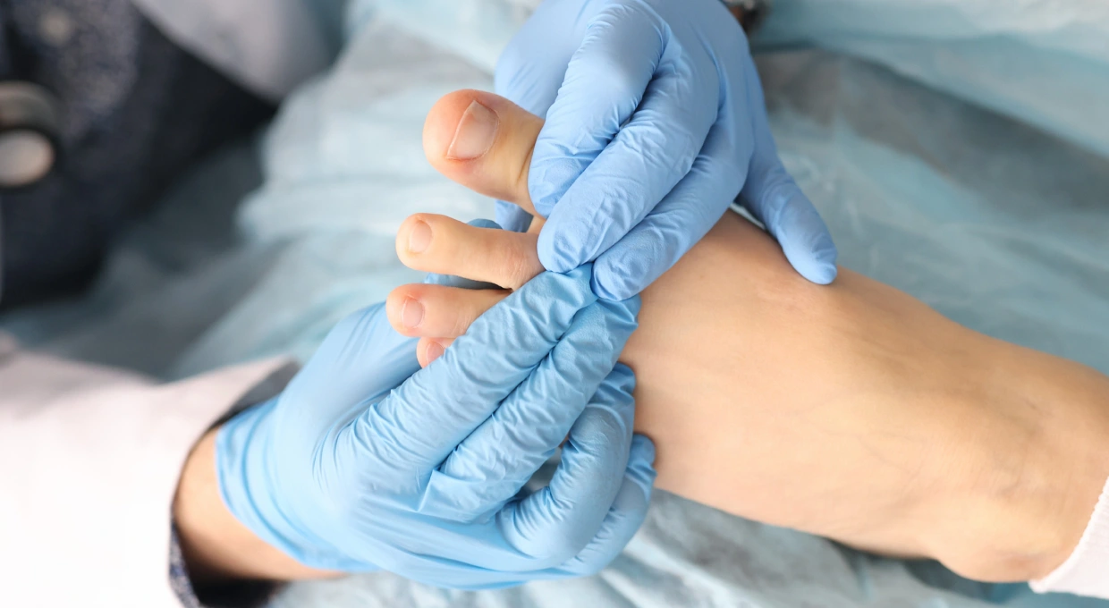
In some cases, a biomechanical study using a pressure platform may be necessary to identify alterations in load distribution during gait and to determine whether customized insoles will be necessary in the postoperative period.
With all this information, the surgeon designs a personalized surgical plan, explaining in detail the proposed technique, the planned osteotomies, whether or not internal fixation (screws) will be used, the specific risks of the case, the expected recovery process and the therapeutic alternatives available. The patient must sign an informed consent form after fully understanding these aspects.
Intervention Day
Preoperative preparation includes the preoperative blood test performed days before, review of the usual medication (especially anticoagulants or antiplatelet medications that may require adjustment), and preoperative fasting according to the anesthetic protocol of the center, typically 6-8 hours for solid foods and 2 hours for clear liquids.
The anesthesia usually used is a regional block of the foot by infiltration of local anesthetic in the nerves that provide sensitivity to the foot. This block is complemented by conscious sedation, which keeps the patient relaxed and comfortable without the need for general anesthesia. This anesthetic approach allows the patient to breathe autonomously, reduces risks associated with general anesthesia and facilitates faster recovery after surgery.
The surgical technique begins with skin marking of anatomical landmarks and planned incision points. Under fluoroscopic control, the surgeon verifies the preoperative planning and proceeds to make the small skin incisions of 2-5 millimeters. Through these minimal portals, he inserts specialized instruments: small caliber motorized drills that allow bone cuts (osteotomies) to be performed with millimetric precision.
Throughout the procedure, the fluoroscope provides real-time images that guide each movement. The surgeon performs the planned osteotomies, mobilizes the bone fragments to correct the deformity and verifies radiologically that the correction achieved is the desired one. This continuous radiological control is one of the keys to the technique, compensating for the lack of direct visualization of the structures.
Once the correction is complete, the small incisions are closed, often with a single stitch or even with surgical adhesive in the smallest incisions. A compressive functional bandage is applied, which serves multiple purposes: it controls edema, maintains a certain compression on the osteotomies favoring their stability and protects the small wounds. Finally, specialized post-surgical footwear is placed and the patient is instructed in its correct use.
The length of the procedure varies depending on the complexity of the case. A single foot procedure with hallux valgus correction by simple osteotomy typically requires 40-60 minutes. More complex cases, involving adjacent toe correction or internal fixation requirements, may extend up to 90 minutes. Bilateral intervention (both feet) in the same surgical procedure, when performed, typically takes 70-90 minutes.
The First Postoperative Hours
After the procedure, the patient is transferred to the recovery room where he/she remains under observation for 1-2 hours. During this time, anesthesia recovery is monitored, the state of the bandage is controlled, it is verified that there is no significant bleeding and oral analgesia is started. It is normal to experience tingling or a “numb foot” sensation during the first hours due to the residual effect of the anesthetic block.
Once the medical team verifies that the patient is stable, can walk with the aid of post-surgical footwear and understands the postoperative instructions, the patient is discharged home. The patient receives written documentation with the care guidelines, the prescribed medication (analgesics, anti-inflammatory drugs, gastric protection, and occasionally antithrombotic prophylaxis depending on the case), the alarm signs that should cause urgent consultation (severe uncontrolled pain, fever, significant bleeding, loss of sensation) and the date of the first check-up.
Recovery: Realistic Expectations by Phases.
Recovery after percutaneous surgery follows a relatively predictable pattern, although with significant individual variations depending on the specific technique employed, the severity of the deformity corrected, the patient’s bone quality and adherence to the postoperative protocol.
The First Two Weeks: Acute Inflammatory Phase
During this period, the main objective is to control pain and edema while allowing the bone consolidation process to begin. Relative rest with the foot elevated above the level of the heart is essential. This elevation, which should be maintained most of the day during the first week, facilitates venous return and significantly reduces swelling.
Cryotherapy (application of cold) is very effective in controlling both pain and edema. It is recommended to apply ice wrapped in a cloth for 15-20 minutes every 2-3 hours while awake. It is important not to apply the cold directly to the skin to avoid skin injury.
Pain during this phase is usually moderate and responds well to conventional analgesics such as paracetamol and non-steroidal anti-inflammatory drugs (if there are no contraindications). Pain intensity is typically greatest during the first 48-72 hours and decreases progressively. If the pain intensifies or does not respond to the prescribed medication, the medical team should be consulted.
Ambulation should be limited to what is strictly necessary: bathing, meals, physiological needs. Post-surgical footwear must be used for any movement, even in the home. During the first few days, canes or crutches may be useful for greater safety, especially in people with balance problems or who live alone.
The initial dressing should not be removed or wet. Some minimal bleeding staining is normal for the first 24-48 hours. The first medical check-up is typically scheduled between days 5-7 postoperatively, at which time the initial dressing is removed, the wounds are checked, the evolution of the edema is evaluated and a new dressing is applied, which the patient will keep until the next check-up.
Weeks 3-6: Initial Consolidation Phase
During this period, the use of post-surgical footwear continues to be mandatory. Although the patient feels more comfortable and may be tempted to try conventional footwear, it is essential to resist this temptation. Bone healing is in process but is not yet sufficiently solid. Premature loading in inappropriate footwear may compromise the correction achieved.
The level of activity can be progressively increased. Short walks inside the home are gradually extended. By the fourth week, many patients can take brief outings outside. However, prolonged walking should be avoided until cleared by the surgeon after verification of radiological consolidation.
Return to work is possible during this phase for sedentary jobs. People who work in an office, telework or activities that allow keeping the foot elevated can generally return between the second and fourth week. Jobs that require prolonged standing require longer periods of sick leave.
The medical check-up between weeks 4-6 is crucial. At this visit, a radiological control is performed to evaluate the degree of bone consolidation achieved. If the images show adequate callus formation and the clinical examination is favorable, the surgeon authorizes the start of the transition to conventional shoes. In some cases, especially if the consolidation appears to be slower, it may be necessary to prolong the use of post-surgical footwear for a few additional weeks.
Physiotherapy may be initiated during this phase if prescribed. Goals include manual lymphatic drainage to reduce residual edema, gentle passive mobilization of the joints to prevent stiffness and, toward the end of this phase, initiation of progressive active mobilization.
Weeks 6-12: Transition and Functional Gain
With medical authorization, the progressive transition to sports shoes is initiated. This change should be gradual: start by wearing sports shoes with a wide toe box and good cushioning for 1-2 hours a day, alternating with post-surgical footwear if discomfort appears. Progressively, over a period of 2-3 weeks, the time spent wearing sports shoes is increased until the post-surgical footwear can be completely dispensed with.
Residual edema is completely normal during this phase. It is common for the foot to remain somewhat swollen, especially at the end of the day after standing or walking. This edema will reduce very gradually over a period of months. The use of soft compression socks may help to control it.
Full return to work is possible for most professions towards the end of this phase. Jobs requiring prolonged standing or continuous travel may require temporary adaptations or reduced working hours initially. Occupations involving significant physical exertion may require longer periods of leave, up to 10-12 weeks.
Driving can generally be resumed between weeks 6-8, provided that it is the left foot that has been operated on (in automatic vehicles) or that sufficient strength and reflexes have been recovered in the right foot. It is essential to consult with the surgeon before resuming driving and to verify with the insurance company that the coverage is maintained.
Physical therapy during this phase intensifies active mobilization exercises, introduces progressive strengthening of the intrinsic musculature of the foot and begins gait re-education through pressure platform analysis if available. The goal is to restore normal gait patterns and prevent compensations that can lead to problems in other structures.
Months 3-6: Advanced Recovery and Return to Sports
During these months, bone healing is complete. The third month radiological control typically shows complete radiological consolidation, with disappearance of the osteotomy line and mature bone formation. This milestone allows for a significant increase in the level of activity.
Everyday footwear can be progressively incorporated. Closed shoes made of flexible leather, moccasins with a good insole and comfortable casual shoes are replacing sneakers. However, it is recommended to maintain healthy footwear criteria: wide toe box, adequate arch support, quality materials and avoid high heels or excessively narrow shoes.
The return to sports should be progressive and supervised. Low impact activities such as swimming can be started as early as 4-6 weeks (once the wounds are completely closed). Exercise cycling is introduced at 6-8 weeks. Hiking on flat terrain can be started at 8-12 weeks. Running requires more caution, generally not before 3-4 months, starting with short sessions of gentle jogging and progressively increasing volume and intensity. Intense impact sports (soccer, basketball, tennis) are typically not fully resumed until 4-6 months postoperatively.
Strengthening and proprioceptive exercises are intensified. Elastic band work to strengthen the foot and ankle musculature, balance exercises on unstable surfaces and toe grip exercises help restore full foot function.
Beyond 6 Months: Consolidating Results
Most patients achieve full recovery by 6 months postoperatively. This means absence of surgery-related pain, complete radiological bone healing, recovery of joint range of motion and ability to perform all daily activities and sports without limitations.
However, some aspects may require more time. Minimal residual edema at the end of the day may persist up to one year postoperatively, especially in patients prone to fluid retention or in warm climates. This residual edema should not be painful or limit activity. Skin sensitivity in the scar area may take up to 12-18 months to completely normalize, with some hypoesthesia (reduced sensitivity) persisting permanently in some cases, although this rarely causes functional problems.
It is essential to maintain good footwear habits over the long term. Although the deformity has been surgically corrected, the predisposing factors (bone structure, ligamentous laxity, gait pattern) persist. The habitual use of narrow shoes or high heels can favor, in the very long term, a recurrence of the deformity. Therefore, it is recommended to reserve less suitable footwear for occasional occasions and to prioritize comfort in everyday life.
Determining if You Are a Suitable Candidate
Not all patients with hallux valgus or foot deformities are ideal candidates for percutaneous surgery. An honest assessment of suitability is critical to avoid frustration and optimize outcomes.
The ideal candidate presents a deformity of mild to moderate severity, documented radiologically with angles in ranges that allow percutaneous correction. They experience pain that significantly limits their daily activities, work or sports practice, having tried unsuccessfully conservative treatment for at least six months. This conservative treatment should have included appropriate footwear, custom insoles if indicated, physical therapy and activity modification. The patient’s bone quality should be adequate, with no severe osteoporosis to compromise healing. It is essential that the patient clearly understands the procedure, the expected results and recovery times, showing willingness to follow the postoperative protocol and the ability to attend the necessary check-ups. The patient’s general state of health must be compatible with a surgical intervention, without decompensated pathologies that significantly increase the surgical risk.
There are situations that require special caution and may constitute relative contraindications. Very severe deformities, with angles that clearly exceed the usual limits of the percutaneous technique, may obtain better results with open techniques that allow wider corrections. The presence of active infections in the foot absolutely contraindicates any elective surgery until complete resolution. Severe peripheral vascular insufficiency compromises healing capacity and dramatically increases the risk of complications, requiring prior vascular assessment and possibly revascularization before considering foot surgery. Decompensated diabetes mellitus, with glycated hemoglobin levels above 8%, is associated with higher rates of infectious complications and delayed healing, and it is advisable to optimize glycemic control prior to surgery. Active smoking significantly increases the risk of healing complications and delayed bone healing; although it does not absolutely contraindicate surgery, smoking cessation at least 4-6 weeks before surgery is strongly recommended. Unrealistic expectations, such as expecting total absence of postoperative discomfort or “immediate” recovery, need to be identified and corrected by adequate information before proceeding with surgery. Uncontrolled severe osteoporosis can compromise bone healing, especially in techniques without internal fixation, and individual assessment and possibly treatment of osteoporosis prior to surgery is necessary.
Comparing Percutaneous Surgery and Open Surgery
The decision between percutaneous and open surgery is not always obvious. Both approaches have strengths and limitations that must be considered in the context of each individual case.
In terms of invasiveness, the difference is clear and objective. Percutaneous surgery uses incisions of 2-5 millimeters that are practically imperceptible once healed, while open surgery requires incisions of 5-10 centimeters that, although they generally heal well, leave a visible linear mark. Soft tissue dissection in open surgery is extensive, requiring sectioning or retraction of muscles, tendons and joint capsules, while the percutaneous technique works between the tissues while respecting their integrity. Intraoperative bleeding in percutaneous techniques rarely exceeds 50 milliliters, while in open surgery it can reach 100-200 milliliters, although these figures do not usually have significant clinical relevance in healthy patients.
Regarding the surgical procedure, percutaneous surgery typically requires 40-60 minutes for one foot, similar in duration to open techniques (60-90 minutes). The main difference lies in the type of anesthesia: percutaneous surgery is generally performed with local block and conscious sedation, allowing same-day discharge, while open surgery most often employs spinal or general anesthesia, although it is also usually outpatient. The learning curve for percutaneous surgery is steeper than for open technique, requiring specific training and accumulated experience to master the technique.
The immediate postoperative period presents significant differences. Pain after percutaneous surgery tends to be mild-moderate and well controlled with standard analgesia, while after open surgery it tends to be moderate-intense, occasionally requiring stronger analgesics. Edema is generally smaller and more localized after percutaneous technique, while in open surgery it tends to be more extensive and prolonged. Ambulation is initiated from the first day in both techniques, although with specialized post-surgical footwear.
The recovery period shows differences that, although present, are less dramatic than some commercial discourse suggests. Return to work for sedentary work is possible around 2-4 weeks after percutaneous surgery, whereas after open surgery it typically requires 4-6 weeks. For standing work, the times are 6-8 weeks and 8-12 weeks respectively. Physiotherapy is initiated around 3-4 weeks in both cases, although the intensity may progress more rapidly after percutaneous technique. Full sports recovery is achieved at 3-6 months after percutaneous and 4-8 months after open, although these figures have wide individual variations.
Regarding medium to long-term results, the available scientific evidence suggests similar results between both techniques in terms of angular correction achieved, long-term patient satisfaction and recurrence rates. Series with 5-year follow-up show satisfaction rates of 85-95% for both approaches. Recurrence rates are 3-7% for percutaneous surgery and 5-10% for open surgery, differences that are not statistically significant in most studies.
Indications differ significantly. Percutaneous surgery is optimal for mild to moderate deformities, in patients with good bone quality, without significant instability of proximal structures. Open surgery has a broader spectrum of indications, being able to address severe deformities, complex joint instabilities, cases of surgical revision and situations where extensive release of contractured soft tissue is required.
Finally, the direct cost of both procedures is similar. Although percutaneous surgery requires specific equipment (high-quality fluoroscopy), the lower need for hospitalization and postoperative resources offsets this cost. In the Spanish public health system, both techniques are covered when there is a clear medical indication, the specific technique used depending on the protocol of the center and the experience of the assigned surgeon.
Frequently Asked Questions
On pain: “Does percutaneous surgery hurt?” During the procedure, local anesthesia and sedation ensure that you will not experience pain. In the hours that follow, as the anesthetic wears off, you will begin to notice discomfort that typically peaks around 24-48 hours postoperatively. This pain is usually mild-moderate in intensity, describable as dull discomfort or a sensation of pressure, rarely as severe sharp pain. The standard analgesics prescribed (combination of paracetamol and anti-inflammatory drugs) usually control it adequately. If the pain is severe, does not respond to medication or intensifies after the first few days, the medical team should be consulted immediately, as it could indicate complications such as compartment syndrome or infection.
On initial mobility: “When will I be able to walk?” Ambulation with post-surgical footwear can be initiated as early as the first postoperative day. However, “being able to walk” does not mean that you must walk extensively. During the first two weeks, ambulation should be limited to what is strictly necessary: movements within the home for bathing, meals and basic needs. The aim of this period is to allow bone healing to begin under optimal conditions, with the foot in relative unloading. The temptation to test the ability to walk or make unnecessary outings should be resisted: although you could probably do so without immediate acute pain, you would be compromising the consolidation process which is in its most critical phases.
On employment: “When do I return to work?” This is perhaps the question with the greatest variability in its answer. A programmer who can telework with the foot elevated might return to work in 1-2 weeks. An office clerk who can keep the foot elevated under the desk frequently may return to work in 2-4 weeks. A salesperson who is required to be on his or her feet serving customers will typically need 6-8 weeks. A construction worker or nurse who spends the entire day on his or her feet and on the move may require 10-12 weeks. Your surgeon will evaluate your specific profession and the physical demands of your job to give you a personalized estimate and, if necessary, propose temporary job accommodations.
About bilateral surgery: “Can I have surgery on both feet simultaneously?” It is technically possible to perform bilateral percutaneous surgery in the same surgical procedure, and some centers offer it. However, most surgeons recommend operating on one foot first and, after full recovery (typically 3-6 months), proceeding with the contralateral foot. The main reason is that operating on both feet simultaneously leaves you with no “healthy” foot to support for the first few weeks, dramatically limiting your mobility and autonomy. This limitation may be manageable for young, healthy patients with excellent family support and who can take a long period of time off work. For most patients, the sequential approach is more practical and safer.
On internal fixation: “Does this surgery use screws?” There is some confusion about this aspect. Some percutaneous surgical techniques achieve stability without the need for internal fixation (screws), by means of a functional dressing that maintains the correction during the first 4-6 critical weeks. Other variants of minimally invasive surgery employ screws, inserted through small incisions under radiological control. Both approaches are “percutaneous” or “minimally invasive” as the incisions remain millimetric. The decision to use screws or not depends on multiple factors: severity of the corrected deformity, bone quality of the patient, specific protocol of the surgeon and surgical philosophy of the center. Both approaches have scientific evidence to support their efficacy. What is essential is that your surgeon clearly explain to you which specific technique will be used in your case and why.
On recurrence: “Can the bunion recur?” This is a legitimate concern. Studies with 5-year follow-up report recurrence rates of 3-7% for percutaneous surgery, figures similar to or slightly lower than those for open surgery. A “recurrence” can be a complete reappearance of the original deformity or, more frequently, a partial deviation less than the initial one. Factors that reduce the risk of recurrence include complete surgical correction of all components of the deformity, good radiologically verified bony healing, subsequent use of appropriate footwear (routinely avoiding narrow shoes or high heels), and control of predisposing factors such as overweight or biomechanical alterations with insoles if indicated. Some risk factors for recurrence (constitutional ligamentous laxity, type of bone structure of the foot) are not modifiable, which explains why even with perfect technique and optimal follow-up of the postoperative protocol, a small percentage of patients experience some degree of recurrence.
On the economic cost: “How much does this surgery cost?” In private practice in Spain, the cost of percutaneous bunion surgery on one foot typically ranges between 2,500-5,000 euros, depending on the center, the complexity of the case and the services included (number of revisions, postoperative physiotherapy, type of anesthesia). Simultaneous bilateral surgery does not double the cost, since it shares operating room and anesthesia expenses, and is generally between 4,000-7,000 euros. Many private medical insurance policies cover this surgery partially or totally when there is a clear medical indication, although it usually requires a waiting period (time from contracting the insurance until it can be used for this coverage) of 6-12 months. In the public health system, bunion surgery is covered when there is a clear indication (limiting pain, documented failure of conservative treatment), although waiting times vary significantly depending on the autonomous community, from 6 to 18-24 months. The specific technique used (percutaneous or open) will depend on the hospital protocol and the experience of the assigned surgeon.
About physiotherapy: “Is physiotherapy mandatory?” It is not strictly mandatory in all cases, but it is highly recommended. Postoperative physiotherapy fulfills multiple objectives that accelerate and optimize recovery. During weeks 3-6, manual lymphatic drainage helps reduce residual edema that hinders mobility and generates discomfort. From week 6 onwards, progressive active and passive mobilization prevents joint stiffness, a frequent complication if the range of motion is not adequately worked on. Between months 2-4, specific muscle strengthening and gait re-education through baropodometric analysis correct compensatory patterns that could generate problems in other structures (knee, hip, back). Your surgeon will assess your evolution and determine whether to prescribe physiotherapy and, if so, how many sessions he/she considers necessary. Some patients with excellent spontaneous evolution and good range of motion may not require it, while others benefit significantly from a structured program of several weeks.
On sporting activity: “Will I be able to do sports again?” Not only will you be able to, but one of the objectives of the surgery is precisely to allow you to resume the activities that the deformity had limited. However, reincorporation must be progressive and supervised. Low-impact activities such as swimming or static cycling can be started relatively soon, around 6-8 weeks. Running requires more caution, typically starting around 3-4 months with very gradual progression of volume and intensity. Sports involving jumping, pivoting or sudden changes of direction (basketball, soccer, tennis) generally wait until 4-6 months. These figures are indicative; your surgeon will schedule radiological controls to verify complete consolidation before authorizing significant progressions in sports load. Attempting to accelerate the return to sport before the bone has fully consolidated significantly increases the risk of complications, from discomfort to stress fractures or loss of the correction achieved.
Common Myths to Clear Up
Marketing and informal information frequently generate misconceptions about percutaneous surgery that should be dismantled with evidence and honesty.
“It’s laser surgery” – This is probably the most widespread myth. There is no laser involved in percutaneous foot surgery. Bone cuts are made with small caliber motorized drills, very similar in principle to those used in open surgery but miniaturized and designed to work through small portals. The medical laser has applications in other fields (ophthalmology, dermatology), but not in bone surgery of the foot.
“No scarring” – Although scars are minimal (2-5 mm) and generally very inconspicuous, they do exist. With time and sun exposure, these small marks can become virtually unnoticeable, but to say that there are no scars is technically incorrect. It is correct to say that the scars are millimetric and much less visible than those of open surgery.
“Recovery is immediate” – This myth generates false expectations and subsequent frustrations. Although recovery is generally faster than with open surgery, it is still a process that requires weeks for initial healing and months for full recovery. You will be able to walk from day one, yes, but with special footwear and limitations. Bone healing requires biological time regardless of the technique used.
“Absolutely no pain” – Postoperative pain is less than in open surgery, but it is not zero. The first 48-72 hours usually involve discomfort that requires analgesia. The intensity is variable among patients, but describing the postoperative period as completely painless is misleading. The honest thing to say is that pain is usually mild-moderate and well controllable with standard medication.
“It fits any bunion, no matter how big” – Severe deformities often require open or mixed techniques that allow for more extensive corrections and more robust stabilization. Percutaneous surgery has technical limitations that must be recognized. An honest surgeon will assess your specific case and tell you clearly whether your deformity is a candidate for percutaneous technique or would require a better result with another approach.
“It’s a new experimental technique” – Percutaneous techniques on the foot were initially developed in the 1980s-90s. What is relatively recent is optimization using high-quality digital fluoroscopy and specifically designed instrumentation. But the concept of performing osteotomies through small incisions is neither new nor experimental. There is ample published scientific literature with follow-ups of 5-10 years supporting its efficacy and safety in appropriately selected cases.
“Insurance does not cover it” – Both the public system and most private insurances cover bunion surgery when there is a clear medical indication. What may vary is the specific technique used by the assigned surgeon, depending on his or her training and the center’s protocols. But the coverage of the procedure itself is not usually a problem.
“It costs much more than normal surgery” – The cost is similar to that of open surgery. Although specific equipment (fluoroscope) is required, hospitalization and postoperative care costs tend to be lower, offsetting the cost of the equipment.
Choosing the Right Professional
The choice of surgeon is probably the most important decision in your process. Some key aspects to consider:
A surgeon qualified to perform percutaneous foot surgery must have specific accredited training in minimally invasive techniques, and general training in foot surgery is not sufficient. These techniques require specific courses, training in reference centers and initial supervised experience. Adequate case volume is essential to maintain proficiency; surgeons who perform these techniques occasionally (less than 20-30 cases per year) may have less predictable results than those with higher volumes. Accumulated experience matters significantly, especially considering the learning curve of these techniques. Asking how many years you have been performing percutaneous surgery and approximately how many cases you have performed is entirely appropriate. Membership in scientific societies specializing in foot and ankle surgery suggests commitment to continuing education and updating knowledge.
Transparent communication is essential. A good surgeon will spend sufficient time explaining the specific technique he/she will use in your case, show examples of previous similar cases (preoperative and postoperative photographs, x-rays), clearly explain the possible risks and complications without minimizing them, discuss therapeutic alternatives if they exist, and answer all your questions without haste or evasiveness. If at any time you feel pressure to decide quickly without time to reflect, or if the answers are vague or evasive, consider seeking a second opinion.
The available equipment is critical in percutaneous surgery. The operating room should have a quality fluoroscope (digital C-arm) that allows high resolution real time images. Specific instruments for minimally invasive foot surgery (drills, special scalpels, mobilization instruments) must be available. Questions on these aspects are fully appropriate.
The follow-up protocol should be clearly structured: how many check-ups are scheduled, at what times, what they include (examination, X-rays, dressing changes), whether physiotherapy is available at the center itself or referral to collaborating physiotherapists with experience in this type of surgery, and how to contact the medical team in case of doubts or problems between check-ups.
Finally, honest individualized assessment is a sign of professional quality. An experienced and ethical surgeon will not try to “sell” the percutaneous technique to all patients. If after the evaluation he/she tells you that your case is not ideal for this technique and proposes an alternative, you are probably dealing with a professional who prioritizes your results over commercial considerations. The honesty of saying “your case would require better results with another technique” is a sign of professional excellence.
Preparing Optimally for the Intervention
Good preoperative preparation contributes significantly to the success of the procedure and a smooth recovery.
In the 2-4 weeks prior to surgery, you must complete all the preoperative studies requested by your surgeon: blood tests including hemogram, coagulation and basic biochemistry; electrocardiogram if you are over 50 years of age or have cardiovascular risk factors; chest x-ray if required for anesthesia. If you are taking anticoagulant or antiplatelet medication (aspirin, clopidogrel, warfarin, direct anticoagulants), you will need coordination between your surgeon and the prescribing physician to assess whether it is necessary to temporarily suspend these drugs. Purchase post-surgical footwear if it is not included in the price of the operation; your surgeon will indicate the specific model and the necessary size.
Prepare your home with initial mobility limitations in mind. If you live in an apartment without an elevator, consider temporary alternatives or at least minimize the frequency of going up/down stairs during the first week. Organize the bedroom with everything you need (water, medication, cell phone, TV remote control) within reach from the bed without having to get up. If you live alone, it is highly recommended to have household or family help for at least the first 3-4 days. Prepare food in advance or organize how you will deal with this aspect the first few days.
In the previous week, stop taking anti-inflammatory drugs (ibuprofen, naproxen) because they increase the risk of bleeding; if you need painkillers, use paracetamol. Do not apply nail polish on the toenails; the nails must be natural to allow coloring and perfusion to be assessed during the postoperative period. Intensify foot hygiene, especially in the interdigital spaces.
The day before, check that you have prepared everything you need for the following day: complete medical documentation (reports, X-rays, blood tests), health card or private insurance card, regular medication that you take regularly, comfortable and loose clothing for the day of surgery (avoid tight pants), clean cotton socks. Check the fasting instructions you have been given, typically 6-8 hours for solids and 2 hours for clear liquids. Do not drink alcohol in the 24 hours prior to surgery.
On the day of surgery, shower with special attention to foot hygiene. Do not apply any creams or lotions to your feet that day. Wear comfortable clothes that you can easily put on without having to bend over excessively. Be sure to bring someone with you; you will not be able to drive after the procedure and it is advisable to have someone with you for the first few hours at home. Leave jewelry and valuables at home. Take your cell phone fully charged.
Conclusion: A Valuable Tool, Not a Panacea
Percutaneous foot surgery represents a genuine and significant advance in the treatment of forefoot deformities. Its advantages in terms of invasiveness, postoperative pain and functional recovery are real and supported by solid scientific evidence. For the right patient, with the right deformity, performed by a surgeon with specific training and experience, it can offer excellent results with minimal surgical trauma.
However, it is not a magical or universal solution. Recognizing its limitations is as important as knowing its advantages. Not all deformities are candidates for this technique. The learning curve is real and the results improve with the surgeon’s experience. Recovery times, although generally more favorable than with open surgery, still require weeks for initial consolidation and months for full recovery.
The most important thing is an honest individualized evaluation by a qualified professional who will assess your specific case, discuss with you all available options (conservative and surgical, percutaneous and open), explain clearly what you can expect in terms of process, results and timing, and allow you to make an informed decision without commercial pressure or false promises.
If you are considering surgery for your hallux valgus or foot deformity, seek that professional and honest assessment. Ask questions, question, seek second opinions if you have doubts. Your decision should be based on complete and realistic information, not on expectations inflated by marketing or anecdotal testimonials.
Professional Consultation in Alicante
At Clínica San Román we perform exhaustive assessments of each case to determine the most appropriate approach according to the individual characteristics of each patient. Our experience of more than 40 years in foot surgery, combined with specific training in minimally invasive techniques, allows us to offer objective advice based on scientific evidence.
First assessment consultation available for a detailed analysis of your case.
Medical Disclaimer: This article is for informational and educational purposes only. It does not constitute personalized medical advice and is not a substitute for face-to-face consultation with a qualified specialist. Recovery times, results and risks vary significantly depending on individual patient characteristics, severity of deformity and specific technique employed. All treatment decisions should be made after individualized medical evaluation.
