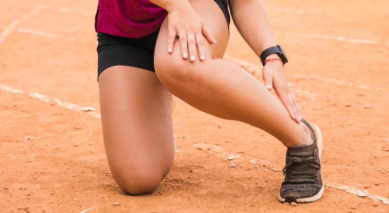
Achilles tendon rupture is one of the most disabling injuries for any active person. It occurs when the tendon that connects the calf muscles to the heel tears in whole or in part, causing severe pain and immediate loss of strength in the ankle. Although it is often associated with athletes, it can affect any individual who makes a sudden effort, a jump or even a misstep. In this article you will find detailed and thorough information on the causes, symptoms, diagnosis and treatment options available, as well as rehabilitation guidelines that will get you back to walking safely.
Functional anatomy of the Achilles tendon
The Achilles tendon is formed by highly resistant collagen fibers that transmit the force generated by the gastrocnemius and soleus muscles to the calcaneus. This structure supports loads of up to twelve times body weight during running and acts as a spring in the swing phase of the stride. The vascularization of the middle third of the tendon, however, is limited, which reduces its capacity for self-repair and explains the frequency of injuries in that specific area.

Causes and risk factors
The rupture is usually caused by a sudden increase in tension on a previously weakened tendon. Common risk factors include:
- Chronic overuse: intense training without adequate rest.
- Sudden changes in activity: going from sedentary life to vigorous exercise.
- Inadequate footwear: stiff soles or lack of cushioning.
- Prolonged intake of corticosteroids or fluoroquinolone antibiotics.
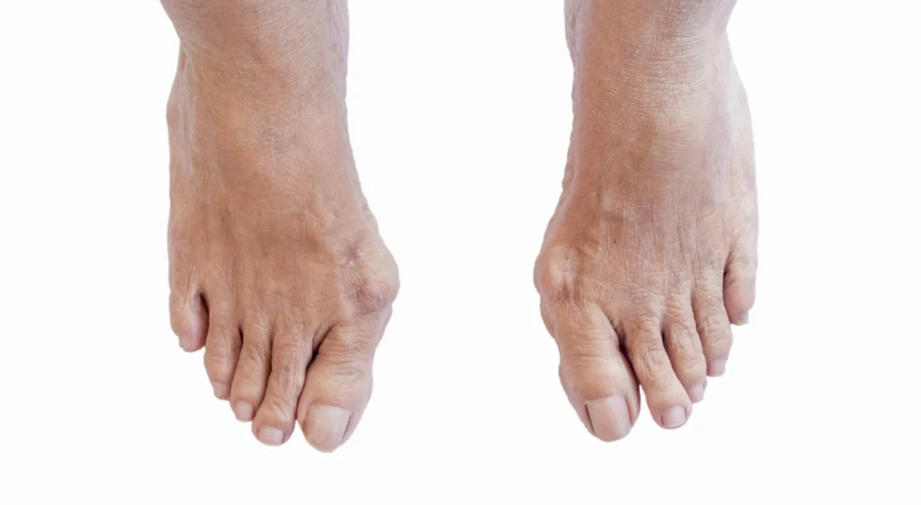
- Biomechanical alterations: pronator foot, previous tendinosis or tendon shortening.
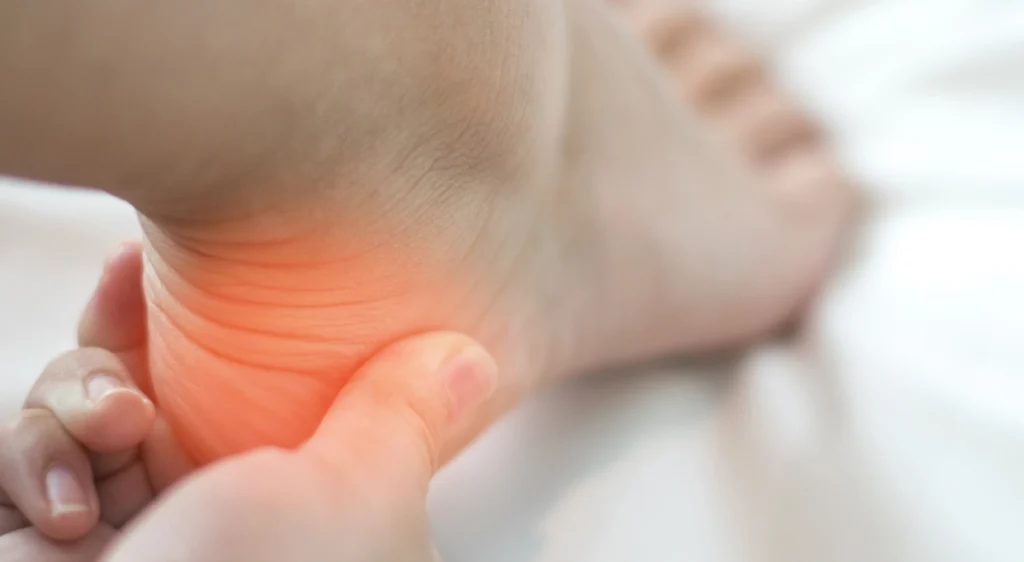
Characteristic symptoms
Achilles tendon rupture manifests itself with an audible snapping sound, described by many patients as the sensation of receiving a stone. This sound is followed by a sharp pain in the back of the ankle and the inability to stand on tiptoe. The heel loses its usual contour and there is a palpable depression in the injured area. The swelling and ecchymosis may spread to the calf in the hours that follow.
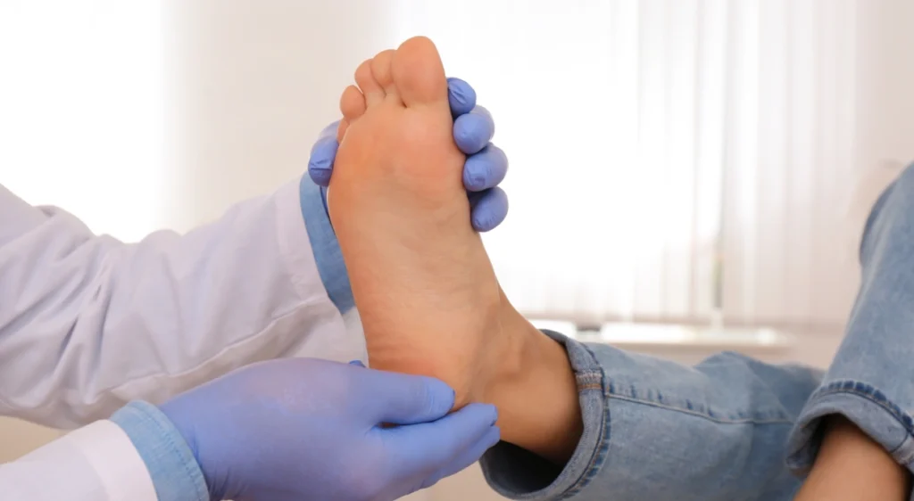
Clinical diagnosis and complementary tests
The first step is a physical examination by a specialist. Thompson’s test, which consists of compressing the calf and observing plantar flexion of the foot, is positive when there is no movement. To confirm the extent and location of the rupture, musculoskeletal ultrasound or magnetic resonance imaging are used to assess the distance between tendon ends and the quality of the tissue, which are essential data for treatment planning.
Treatment options
Conservative treatment
Indicated in partial breaks or in patients of low sport level. The ankle is immobilized in equinus (plantar flexion) by means of orthopedic boot or splint during the first three weeks. Subsequently, the angle of the splint is adjusted to gradually bring the tendon edges closer together, while guided isometric exercises are initiated. Success depends on the correct approximation of the strands and strict physiotherapy.
Surgical treatment
Recommended for complete ruptures and for people who wish to recover high levels of activity. There are two main techniques:
- Classic open suture: allows direct vision and reinforcement with grafts, but with a higher risk of skin complications.
- Minimally invasive suture (percutaneous or endoscopic): reduces scarring and risk of infection, facilitates early functional recovery.
The choice is based on the length of the tear, the quality of the tissue and the patient’s functional goals.
Rehabilitation and recovery times
Supervised physical therapy is key to restoring tendon elasticity and strength. The standard protocol includes:
- Protective phase (0-3 weeks): immobilization in equine, partial loading with crutches.
- Controlled mobility phase (3-6 weeks): progressive removal of heel pad, passive and active assisted joint range-of-motion exercises.
- Strengthening phase (6-12 weeks): concentric and eccentric calf work, proprioception in stable plane.
- Functional reintegration phase (12-24 weeks): light running, plyometric jumps and monopodal standing.
Return to high-demand sports activities ranges from six to nine months, depending on adherence to the program and biological healing.
Potential complications
The most common are rerupture, scar adhesion and ankle stiffness. Superficial infections or deep vein thrombosis are less frequent, but require monitoring. Periodic follow-up with ultrasound allows detection of tendon weaknesses and adaptation of the training load.
Prevention strategies
- Plan for increases in exercise volume and intensity of up to 10% per week.
- Perform daily calf and plantar fascia stretches.
- Use shoes with adequate cushioning and moderate heel cushioning.
- Strengthen the posterior chain through eccentric exercises (heel step down).
- Correct biomechanical imbalances with customized insoles if recommended by the specialist.
When to see a specialist
If you experience sudden pain in the back of the ankle, feel a snapping sound when trying to push off, or find it impossible to stand on tiptoe, you should see a podiatrist or orthopedic surgeon immediately. Early diagnosis and treatment significantly improve the prognosis and reduce the risk of sequelae.
Conclusion
Achilles tendon rupture is a serious injury, but it is treatable with excellent results when early intervention and a rigorous rehabilitation plan are followed. Understanding the risk factors, recognizing the symptoms and acting quickly are the cornerstones that make the difference between a full recovery and the development of chronic complications.
Have you suffered an Achilles tendon injury or are you experiencing persistent heel pain? At Clínica San Román we have specialists in foot surgery and advanced physiotherapy ready to provide you with an accurate diagnosis and a safe recovery. Complete our contact form to book your appointment. Your step forward begins today.